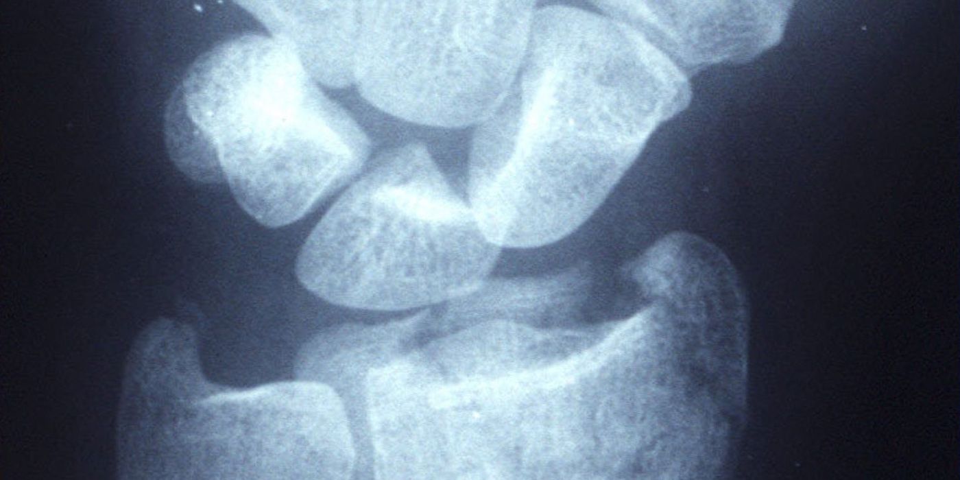
A distal radius fracture is a break of the radius at the wrist, most often caused by a fall onto an outstretched hand. It is the most common fracture seen in UK emergency departments, with a bimodal distribution: high-energy injuries in young adults and fragility fractures in older patients, predominantly post-menopausal women. Most are managed in a cast; surgery is reserved for unstable or significantly displaced fractures, guided in the UK by the December 2017 BOAST on Distal Radial Fractures and NICE guideline NG38 (Fractures: non-complex).
How do distal radius fractures present and how are they diagnosed?
Patients present with wrist pain, swelling and often a visible deformity — classically the "dinner fork" of a dorsally displaced (Colles-type) fracture. Volarly displaced (Smith-type) and volar shearing (Barton-type) patterns are less common but important because they behave differently. On first assessment, do the following:
- Document the mechanism of injury and clinical findings, including skin integrity, circulation and sensation at presentation — an explicit requirement of the Dec 2017 BOAST. Re-examining and recording median nerve sensation (radial three and a half digits) after any manipulation is good practice, reinforced by the Dec 2021 Peripheral Nerve Injury BOAST.
- Obtain posteroanterior and lateral radiographs centred at the wrist. CT is reserved for complex intra-articular patterns or surgical planning.
- Examine for associated injuries: distal ulnar fractures, distal radioulnar joint (DRUJ) disruption, scaphoid tenderness and elbow injury.
What is the initial management?
Open fractures are managed along the national open fracture pathway (combined BOA/BAPRAS standards): urgent assessment, antibiotics, and combined orthoplastic surgical care. For the common closed injury:
- Manipulate displaced fractures under appropriate anaesthesia. The Dec 2017 BOAST states that if manipulation is indicated "it should be undertaken using regional anaesthesia, performed by a suitably qualified and trained practitioner". NICE NG38 recommends considering intravenous regional anaesthesia (Bier's block) for dorsally displaced fractures in adults (16 or over) and specifically advises against manipulating under nitrous oxide alone — a frequent audit failure in UK departments.
- Apply a well-moulded cast with the wrist in neutral flexion and three-point moulding; extreme palmar flexion (the historical "Cotton–Loder" position) risks median nerve compression and stiffness and should be avoided.
- Refer to fracture clinic so the patient is assessed within 72 hours, as per the BOAST.
- Re-image unstable patterns at 1–2 weeks to detect re-displacement while corrective surgery is still straightforward. Stable, minimally displaced fractures can move early: the BOAST advises considering early mobilisation from a removable support once pain allows, and routine radiographs at cast removal are not required unless there is clinical concern.
Who needs surgery? Acceptable radiographic parameters
Neither the BOAST nor NG38 prescribes exact numerical thresholds; the decision weighs alignment against the patient's age, function and demand. The parameters below are the widely used working criteria in UK practice (good practice, not verbatim guideline figures):
| Parameter | Normal value | Commonly accepted limit |
|---|---|---|
| Radial inclination | ~22° | Loss of >5° suggests instability |
| Radial height | ~11 mm | Shortening >2–3 mm (or significant positive ulnar variance) |
| Volar tilt | ~11° volar | Dorsal angulation beyond ~10° from neutral |
| Articular step | Congruent | Step or gap >2 mm |
The BOAST frames the decision by age group:
- Patients over about 65: non-operative management is acceptable for dorsally displaced fractures without significant deformity or neurological compromise. Radiographic malalignment correlates poorly with function in lower-demand older patients, and this is a deliberate, evidence-based tolerance — not neglect. Fragility-fracture patients should also be assessed for falls risk and bone health (fracture liaison service referral is a BOAST standard in its own right).
- Younger patients: assess ulnar variance, intra-articular step and dorsal tilt against the individual's functional demands; thresholds are applied more strictly because malunion is more likely to matter.
- Volarly displaced fractures at any age are inherently unstable and, per the BOAST, "should be considered for open reduction and plate fixation" — a cast cannot reliably hold them.
K-wires, plates or a cast — what does the evidence say?
This is the classic ST3 interview territory, and the UK position is unusually well defined because of two large NIHR trials led by Costa and colleagues.
DRAFFT (BMJ 2014): K-wires vs volar locking plates
461 adults with dorsally displaced distal radius fractures (reducible closed, articular surface reducible) were randomised to percutaneous K-wire fixation or volar locking-plate fixation. There was no clinically relevant difference in the Patient-Rated Wrist Evaluation (PRWE) at 3, 6 or 12 months (difference at 12 months −1.3, 95% CI −4.5 to 1.8), no difference in complications, and K-wiring was substantially cheaper (mean saving around £727 per patient) and quicker. Five-year follow-up (Costa ML et al., Bone Joint J 2019;101-B(8):978–983) showed no late divergence. This evidence underpins the NG38 recommendation to "offer K-wire fixation if no fracture of the articular surface of the radial carpal joint is detected, or displacement of the radial carpal joint can be reduced by closed manipulation", reserving open reduction and internal fixation for fractures that cannot be reduced closed.
DRAFFT2 (BMJ 2022): K-wires vs a moulded cast
The follow-on question was whether, once a fracture is manipulated, wires add anything over a properly moulded cast. 500 adults (aged 16 and over) were randomised after manipulation to K-wire fixation or a moulded cast. At 12 months there was no significant difference in PRWE (adjusted mean difference −0.34, 95% CI −4.33 to 3.66; P=0.87). The crucial caveat: 33 of 255 patients (13%) in the cast group lost reduction and required surgical fixation in the first 6 weeks — so casting is a reasonable first-line strategy only with mandatory early radiographic surveillance and a low threshold for delayed fixation.
Timing of surgery
When fixation is needed, NG38 and the BOAST align: operate within 72 hours of injury for intra-articular fractures and within 7 days for extra-articular fractures; when a fracture re-displaces in a cast, fixation should follow within 72 hours of the decision to operate.
How should median nerve problems be managed?
Median nerve symptoms are the key neurological issue at the wrist. Distinguish three situations:
- Acute carpal tunnel syndrome — progressive, severe median nerve pain and deficit after injury or manipulation. This is a surgical emergency: urgent reduction of the fracture and, if the deficit persists or progresses, emergency carpal tunnel decompression, usually with fracture fixation at the same sitting.
- Static, incomplete contusion-type symptoms present from the outset may be observed after reduction, with clear documentation and early senior review; any deterioration mandates decompression. The Dec 2021 BOAST on Peripheral Nerve Injury reinforces structured examination, documentation and escalation of nerve deficits in trauma.
- Iatrogenic risk: avoid extreme palmar-flexed casting, and re-examine and document nerve function after every manipulation.
- Late carpal tunnel syndrome and other complications warrant routine follow-up advice: patients should receive written information on expected recovery and a self-referral route back to the service (a BOAST standard).
What are the complications and prognosis?
- Malunion — the commonest complication; may cause DRUJ dysfunction, reduced grip and midcarpal malalignment. Symptomatic malunion in active patients may need corrective osteotomy.
- Extensor pollicis longus rupture — classically after minimally displaced fractures (attritional/ischaemic at Lister's tubercle); treated with extensor indicis proprius transfer.
- Complex regional pain syndrome, stiffness, pin-site infection (wires), and flexor tendon irritation or rupture from prominent volar plates.
- Carpal tunnel syndrome, early or late, as above.
Most patients regain functional wrist movement by 12 months whichever evidence-based pathway is used; older patients tolerate deformity well, while young, high-demand patients justify a lower threshold for anatomical restoration.
Key points
- Document skin, circulation and sensation (median nerve) at presentation — a Dec 2017 BOAST requirement; re-examine and document after every manipulation (good practice, reinforced by the Dec 2021 Peripheral Nerve Injury BOAST).
- Manipulate under regional anaesthesia (consider Bier's block); never under nitrous oxide alone (NICE NG38).
- Fracture clinic review within 72 hours; re-image unstable patterns at 1–2 weeks.
- Over-65s with dorsal displacement and no significant deformity or nerve compromise: non-operative management is acceptable.
- Volarly displaced fractures are unstable — plan open reduction and volar plate fixation.
- DRAFFT: K-wires equal plates for reducible dorsally displaced fractures, at lower cost. DRAFFT2: a moulded cast equals wires at 12 months, but 13% need delayed fixation — surveillance is mandatory.
- Fix intra-articular fractures within 72 hours, extra-articular within 7 days.
- Acute, progressive median nerve deficit = emergency reduction ± carpal tunnel decompression.
- Every fragility fracture triggers bone health and falls assessment.