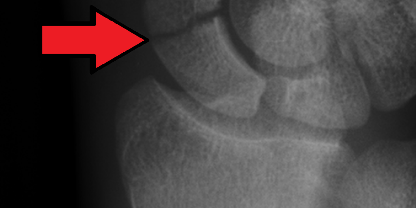
A scaphoid fracture is a break in the scaphoid, the boat-shaped carpal bone on the radial side of the wrist and the most commonly fractured carpal bone, typically caused by a fall onto an outstretched hand in a young adult. Its retrograde, predominantly single-vessel blood supply makes it uniquely vulnerable to nonunion and avascular necrosis, so the priorities are early diagnosis — with MRI if radiographs are normal — and treatment that reliably achieves union. Most undisplaced waist fractures unite in a cast; displaced and proximal pole fractures are usually fixed surgically.
Why is the scaphoid so prone to nonunion?
The scaphoid's vascular anatomy explains almost all of its clinical behaviour. In the classic cadaveric injection study by Gelberman and Menon (Journal of Hand Surgery, 1980), 70–80% of the intraosseous blood supply — and the entire proximal pole — enters through foramina on the dorsal ridge from the dorsal carpal branch of the radial artery, perfusing the bone in a retrograde (distal-to-proximal) direction. The remaining 20–30%, around the distal tuberosity, comes from volar radial artery branches.
- A fracture through the waist can devascularise the proximal fragment, since its only supply crosses the fracture line.
- The more proximal the fracture, the higher the risk of delayed union, nonunion and avascular necrosis (AVN) — proximal pole fractures carry the worst prognosis.
- Around 80% of the scaphoid surface is articular cartilage, leaving little periosteum for callus formation; healing is largely by primary bone healing, which demands stability.
How does a scaphoid fracture present and how is it diagnosed?
The typical patient is a young adult after a fall onto an outstretched, extended wrist, or a punch or handlebar injury. Examine for the classical triad (good practice — sensitive but poorly specific individually, more useful in combination):
- Tenderness in the anatomical snuffbox;
- Tenderness over the scaphoid tubercle (volar);
- Pain on axial compression of the thumb (telescoping).
Request dedicated scaphoid radiographs (PA, lateral, and oblique/ulnar-deviated PA "scaphoid views"). Initial radiographs miss a substantial minority of fractures. This is where UK guidance is explicit: NICE guideline NG38, Fractures (non-complex): assessment and management (2016), recommends: "Consider MRI for first-line imaging in people with suspected scaphoid fractures following a thorough clinical examination." In practice, most UK units image with radiographs first and reserve early MRI for the clinically suspected fracture with normal films; the traditional pathway of two weeks' immobilisation followed by repeat radiographs delays diagnosis, over-treats the majority who have no fracture, and is increasingly replaced by early definitive imaging (MRI, or CT where MRI is unavailable). CT is also the imaging of choice for defining displacement, fracture geometry (e.g. humpback deformity) and union.
How are scaphoid fractures classified?
The Herbert classification is the most quoted in the ST3 interview because it links morphology to stability and therefore to treatment:
| Herbert type | Description | Implication |
|---|---|---|
| A | Stable acute fracture (A1 tubercle, A2 incomplete waist) | Cast treatment; reliable union |
| B | Unstable acute fracture (B1 distal oblique, B2 complete/displaced waist, B3 proximal pole, B4 trans-scaphoid perilunate fracture-dislocation) | Usually operative fixation |
| C | Delayed union | Reassess; consider fixation ± graft |
| D | Established nonunion (D1 fibrous, D2 pseudarthrosis) | Nonunion surgery |
Fractures are conventionally called displaced if there is ≥1 mm translation or step, or angular deformity (e.g. lateral intrascaphoid angle >35°, radiolunate angle >15° suggesting a DISI pattern) — features best assessed on CT.
How are acute scaphoid fractures managed?
Initial management
Immobilise the clinically suspected or confirmed fracture at first presentation (splint or below-elbow cast), provide analgesia, and arrange definitive imaging and fracture-clinic review within days rather than weeks (good practice). There is no evidence that including the thumb in the cast improves union; a standard below-elbow cast leaving the thumb free is acceptable and was the cast used for the large majority of the SWIFFT cast arm (the pragmatic protocol left thumb inclusion to the treating clinician).
Cast or fix? The SWIFFT evidence
For the commonest injury — the undisplaced or minimally displaced (≤2 mm) bicortical waist fracture — the question of early percutaneous/open screw fixation versus cast was answered by the SWIFFT trial (Dias et al, The Lancet, 2020; NIHR HTA 2020), a pragmatic multicentre RCT of 439 adults across 31 UK hospitals randomised to headless compression screw fixation (n=219) or below-elbow cast for 6–10 weeks (n=220), with prompt fixation of any confirmed nonunion in the cast arm:
- No clinically relevant difference in the primary outcome, Patient-Rated Wrist Evaluation at 52 weeks: cast 14.0 vs surgery 11.9; adjusted difference −2.1 (95% CI −5.8 to 1.6; p=0.27), below the 6-point minimal important difference.
- Nonunion at 52 weeks was rare in both arms (surgery 1/203 vs cast 4/205 analysed); 17 cast-arm patients underwent surgery for confirmed nonunion.
- Complications and reoperations were more frequent after primary surgery, and surgery cost around £1,295 more per patient.
The practice message: treat undisplaced and minimally displaced waist fractures in a cast, monitor union actively, and fix confirmed nonunion promptly — "aggressive surveillance, selective fixation". Offer primary fixation where the fracture is displaced >2 mm, comminuted or proximal pole, where there is an associated perilunate injury, or (as a shared decision) in patients who cannot tolerate prolonged casting. Percutaneous headless compression screw fixation along the central scaphoid axis is the standard technique for suitable waist fractures; proximal pole fractures are usually fixed through a dorsal approach.
Trans-scaphoid perilunate fracture-dislocations (Herbert B4) are a distinct emergency: urgent reduction, then open reduction, scaphoid fixation and ligament repair.
How is scaphoid nonunion managed?
Suspect nonunion when pain persists beyond 8–12 weeks or radiographs show cysts, sclerosis or resorption; confirm with CT. Untreated nonunion leads to a predictable degenerative sequence — scaphoid nonunion advanced collapse (SNAC) — beginning at the radial styloid, progressing to the radioscaphoid then midcarpal joints, with humpback collapse and DISI deformity. Management depends on fragment vascularity (MRI ± contrast, or punctate bleeding at surgery), deformity, and whether arthritis is established:
- Stable fibrous nonunion, no deformity: screw fixation, often with cancellous autograft.
- Humpback deformity, viable proximal pole: anterior wedge (corticocancellous, e.g. iliac crest) non-vascularised graft with fixation to restore length and correct DISI.
- Proximal pole AVN or failed previous grafting: vascularised bone graft — the 1,2-intercompartmental supraretinacular artery (Zaidemberg) pedicled graft from the distal radius, or a free medial femoral condyle graft for larger defects.
- Established SNAC wrist (salvage): radial styloidectomy for early disease; scaphoid excision with four-corner fusion or proximal row carpectomy for SNAC II–III; total wrist fusion for pancarpal arthritis; wrist denervation as a motion-preserving adjunct.
What are the complications and prognosis?
- Union: around 90–95% of undisplaced waist fractures unite in a cast; proximal pole and displaced fractures fare substantially worse, which is why they are fixed.
- Nonunion and AVN: the principal complications, driven by the retrograde blood supply; risk rises with proximal location, displacement and delayed diagnosis.
- SNAC arthritis: the long-term consequence of neglected nonunion.
- Surgical complications: SWIFFT showed a meaningful reoperation and complication burden after fixation (screw prominence, infection, CRPS), which routine casting avoids.
- Missed scaphoid fracture remains a recurrent source of litigation in emergency care — a safety-netted imaging pathway matters medicolegally as well as clinically (good practice).
Key points
- The dorsal carpal branch of the radial artery supplies 70–80% of the scaphoid, including the whole proximal pole, retrogradely (Gelberman & Menon, 1980) — the anatomical basis of nonunion and AVN.
- Snuffbox, tubercle and thumb-compression tenderness with normal radiographs still means a suspected scaphoid fracture: NICE NG38 says consider MRI as first-line imaging after thorough clinical examination.
- SWIFFT (Lancet 2020): for waist fractures displaced ≤2 mm, cast with active surveillance equals early screw fixation at 52 weeks, with fewer complications and lower cost — fix confirmed nonunion promptly.
- Fix primarily: displacement >2 mm (many would fix >1 mm), proximal pole fractures, and trans-scaphoid perilunate injuries.
- Nonunion surgery is tailored to vascularity and deformity: fixation ± cancellous graft, wedge graft for humpback, vascularised graft for AVN, and salvage (four-corner fusion or PRC) for SNAC wrist.