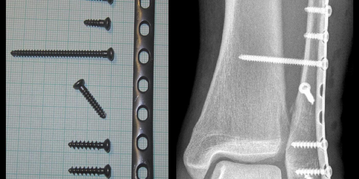
An ankle fracture is a break of the distal fibula, distal tibia (medial or posterior malleolus) or both, usually caused by a twisting injury, and it is one of the commonest injuries treated by trauma and orthopaedic units. The central management question is stability: a stable fracture with a congruent ankle mortise can be treated in a splint or cast with immediate weight-bearing, whereas an unstable fracture that displaces the talus within the mortise usually needs reduction and, in most adults, operative fixation. UK management is defined by the British Orthopaedic Association Standard for Trauma (BOAST) The Management of Ankle Fractures (August 2016).
How are ankle fractures classified?
Two systems dominate, and both are core interview knowledge. The Danis-Weber (AO) classification describes the level of the fibular fracture relative to the syndesmosis, which correlates with the likelihood of syndesmotic injury and instability. The Lauge-Hansen classification describes the position of the foot and the deforming force at the moment of injury, and predicts the sequence of ligamentous and bony failure — useful for understanding occult medial or posterior injuries and for planning closed reduction (reversing the mechanism).
| Weber type | Level of fibular fracture | Syndesmosis / stability | Approximate Lauge-Hansen correlate |
|---|---|---|---|
| A | Below the syndesmosis (infra-syndesmotic) | Syndesmosis intact; usually stable | Supination-adduction |
| B | At the level of the syndesmosis (trans-syndesmotic) | Variable — stability depends on the medial side | Supination-external rotation (the commonest pattern) |
| C | Above the syndesmosis (supra-syndesmotic) | Syndesmosis disrupted; usually unstable | Pronation-external rotation / pronation-abduction |
Neither system dictates treatment by itself. The operative question is whether the talus remains congruent in the mortise under physiological load — a Weber B fracture with an intact deep deltoid ligament is stable and behaves benignly; the same fibular fracture with deltoid failure is an unstable bimalleolar-equivalent injury.
How do ankle fractures present and how is stability assessed?
Patients present after a twisting injury with pain, swelling and inability to weight-bear. As per the August 2016 BOAST, "the mechanism of injury and clinical findings, including skin integrity, assessment of circulation and sensation, should be precisely documented at presentation", and comorbidities that influence treatment — diabetes, neuropathy, peripheral vascular disease, osteoporosis, smoking, alcohol excess — should be documented from the outset.
- Imaging: radiographs should be centred on the ankle and include a true lateral and a mortise view (BOAST). NICE NG38 (Fractures: non-complex, 2016) recommends using the Ottawa ankle and foot rules to determine whether an X-ray is needed in people over 5 years with suspected ankle fractures.
- Look proximally: tenderness over the proximal fibula mandates full-length leg radiographs to exclude a Maisonneuve injury; CT is helpful for complex patterns, particularly posterior malleolar fragments (BOAST).
- Radiographic instability: talar shift or tilt, medial clear space widening greater than the superior clear space (a common working threshold is >4 mm), and tibiofibular clear space widening suggest an incongruent mortise. Medial tenderness or bruising alone is an unreliable predictor of deep deltoid rupture — this is good practice rather than a guideline statement.
- Uncertain stability: the BOAST states that "in fracture patterns where stability is uncertain, patients should be reviewed within 2 weeks with further radiographs (weight bearing if possible) to confirm the position remains acceptable". NICE NG38 corroborates this, recommending orthopaedic follow-up within 2 weeks of a unimalleolar ankle fracture if there is uncertainty about stability. A Weber B fracture that maintains a congruent mortise on standing films can be managed non-operatively; gravity or manual stress views are an alternative but weight-bearing films better reflect physiological load.
What is the initial (emergency department) management?
- Reduce and splint urgently any clinically deformed ankle: "reduction and splinting should be performed urgently for clinically deformed ankles. Radiographs should be obtained before reduction unless this will cause an unacceptable delay" (BOAST, Aug 2016). A fracture-dislocation threatening the skin is an orthopaedic emergency — do not wait for X-ray.
- Re-examine and document neurovascular status after every reduction, and obtain check radiographs confirming an adequate reduction before the patient leaves the emergency department (BOAST).
- Open fractures follow the BOA/BAPRAS Open Fractures BOAST (Dec 2017): intravenous antibiotics as soon as possible and ideally within one hour of injury, debridement within 12 hours for high-energy and 24 hours for low-energy injuries (immediately if agricultural/aquatic/sewage contamination or vascular compromise), and management in an orthoplastic centre with definitive soft-tissue cover within 72 hours.
- Stable fractures "should be treated with analgesia, splinting and patients allowed to bear weight as tolerated. Further follow-up may not be necessary" (BOAST) — an important, frequently examined point: stable Weber A/B injuries do not need routine prolonged immobilisation or serial clinics.
- VTE risk assessment should follow local protocols for any immobilised lower limb (BOAST).
Who needs surgery — ORIF or non-operative management?
The BOAST splits definitive management by age and stability:
- Unstable mortise, under 60 years: "early fixation (on the day or day after injury) is recommended in the majority of patients under 60 years when the ankle mortise is unstable." If the window is missed because of swelling, delayed ORIF once soft tissues allow is standard practice, but early surgery avoids that delay altogether.
- Unstable mortise, over 60 years: "in patients over 60 years close contact casts are an option if reduction can be maintained" — see the elderly evidence below.
- At surgery: achieve anatomical reduction and stable fixation (typically a lateral fibular plate with lag screw, with medial and posterior malleolar fixation as indicated), then "the syndesmosis should then be assessed and stabilised if unstable", with intra-operative radiographs confirming reduction (BOAST).
- Rehabilitation: the BOAST states most patients should be allowed to weight-bear as tolerated in a splint or cast after fixation unless fixation stability, neuropathy or soft-tissue concerns dictate otherwise. This is now supported by the WAX trial (Bretherton et al, The Lancet, 2024): early weight-bearing at two weeks was non-inferior to delayed weight-bearing at six weeks and highly likely to be cost-effective.
- Follow-up: after surgery, patients "should be followed up in fracture clinic within 6 weeks of surgery to detect complications and confirm maintenance of reduction on radiographs" (BOAST), plus written advice on recovery, work and driving.
How should ankle fractures in older patients be managed?
This is the classic evidence question at ST3 interview. Elderly, osteoporotic bone and fragile soft tissues make standard plating riskier, and two UK trials frame the options:
- AIM trial (Willett et al, JAMA, 2016): 620 patients over 60 with unstable ankle fractures randomised to close contact casting (a minimally padded, moulded cast applied under anaesthesia) or ORIF across 24 NHS centres. Ankle function at six months was equivalent, with fewer wound and infection problems in the casting arm and a cost saving of roughly £650 per patient over six months (NHS mean difference −£644); the trade-off was that around one in five casted patients lost reduction and required later surgery. Three-year follow-up (Keene et al, JAMA, 2018) showed no significant functional difference. Close contact casting is therefore a legitimate option in the older patient provided reduction can be achieved and maintained, exactly as the BOAST states — with close radiographic surveillance.
- Fibular nail RCT (White et al, Bone & Joint Journal, 2016): 100 patients over 65 randomised to a percutaneous intramedullary fibular nail or standard ORIF. The nail achieved accurate reduction with a significantly lower rate of soft-tissue complications, comparable radiographic and patient-reported (Olerud-Molander) outcomes at one year, and better cost-effectiveness — making it an attractive option where the skin is compromised or the bone is very osteoporotic.
- Frail patients: the BOAST The Older or Frail Orthopaedic Trauma Patient (2019) applies to fragility fractures generally: surgery in the frail should allow full weight-bearing for daily activities and be performed within 36 hours of admission, with comprehensive geriatric assessment commencing within 72 hours of injury, day-one physiotherapy, and falls and bone-health review. In practice this pushes surgeons towards fixation constructs that tolerate immediate loading (fibular nail, posterior antiglide or locking plates, or a hindfoot nail in the non-ambulant or severely neuropathic patient — the latter being a salvage-type, good-practice option rather than a guideline recommendation).
- Diabetes and neuropathy deserve explicit mention: they multiply wound, infection and Charcot-type failure risks, argue for more robust fixation and longer protected immobilisation, and are precisely the comorbidities the BOAST insists are documented at presentation.
How is the syndesmosis assessed and managed?
The distal tibiofibular syndesmosis (anterior-inferior and posterior-inferior tibiofibular ligaments, interosseous ligament/membrane) maintains the fibula in the tibial incisura. Suspect injury in Weber C and Maisonneuve patterns, and test it in every operated ankle: after malleolar fixation, perform an intra-operative stress examination (lateral fibular pull with a bone hook — the Cotton test — and/or an external rotation stress view under fluoroscopy). The BOAST is explicit: assess the syndesmosis at surgery and stabilise it if unstable, confirming reduction radiographically. Fixation is with syndesmotic screws or a suture-button device; malreduction is the key determinant of poor outcome, so many surgeons reduce under direct vision or check with intra-operative imaging or CT (good practice). A fixed posterior malleolar fragment often restores syndesmotic stability via the posterior-inferior tibiofibular ligament, avoiding the need for a separate syndesmotic device. There is no universally agreed protocol for screw removal; routine removal is not mandatory.
What are the complications and prognosis?
- Early: wound breakdown and surgical site infection (highest risk in diabetes, neuropathy, smokers and the elderly), venous thromboembolism, and loss of reduction in cast-treated unstable fractures.
- Late: malunion and syndesmotic malreduction (the strongest modifiable predictors of post-traumatic osteoarthritis), stiffness, prominent or failed metalwork, complex regional pain syndrome, and non-union (uncommon, mainly medial malleolus).
- Prognosis: anatomically reduced, congruent ankles do well; most patients regain functional mobility over 6–12 weeks, though swelling and stiffness commonly persist for a year. Talar shift left unreduced dramatically reduces tibiotalar contact area and accelerates arthritis — the biomechanical rationale (from Ramsey and Hamilton's classic contact-area work) for insisting on a congruent mortise.
Key points
- Stability — not the Weber letter — decides treatment: a congruent mortise under load can be treated non-operatively with immediate weight-bearing.
- Deformed ankles are reduced and splinted urgently, with documented neurovascular reassessment and check films before leaving the ED (BOAST, Aug 2016).
- Uncertain stability: review within 2 weeks with further radiographs — weight bearing if possible (BOAST; NICE NG38 similarly advises orthopaedic follow-up within 2 weeks).
- Unstable mortise under 60: fix early, on the day of or day after injury (BOAST).
- Over 60: close contact casting is evidence-based (AIM, JAMA 2016) if reduction holds; the fibular nail gave significantly fewer wound infections than ORIF in over-65s (White, Bone & Joint J 2016).
- Assess the syndesmosis in every operated ankle and stabilise if unstable; malreduction drives poor outcomes.
- Allow most patients to weight-bear as tolerated after fixation (BOAST; WAX trial, Lancet 2024).