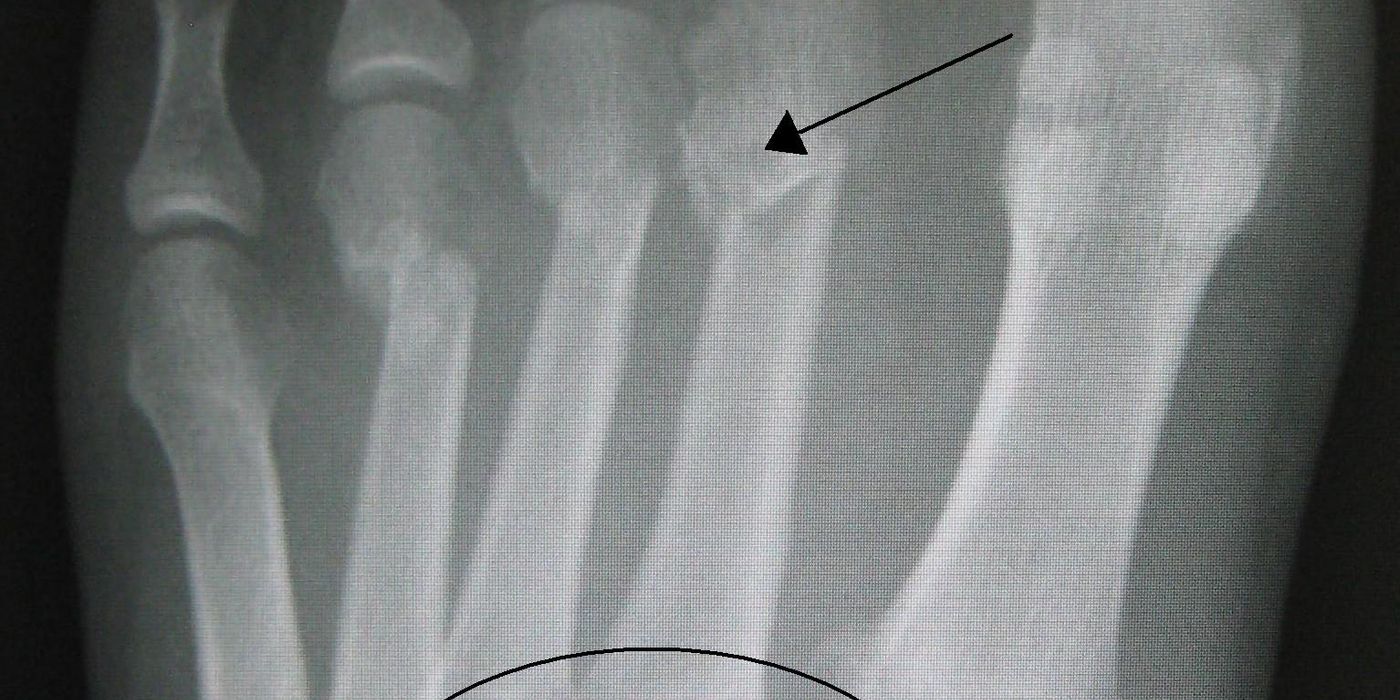
A Lisfranc injury is a disruption of the tarsometatarsal (TMT) joint complex of the midfoot, ranging from a subtle ligamentous sprain to a grossly displaced fracture-dislocation. It is easily missed — a figure of up to 20% of injuries being overlooked or misdiagnosed at first presentation is widely reported — and a missed unstable injury leads to arch collapse and disabling post-traumatic arthritis. Unstable injuries require anatomical reduction with stable internal fixation or, particularly for purely ligamentous patterns, primary arthrodesis.
What is the Lisfranc joint and why does its anatomy matter?
The Lisfranc joint complex comprises the articulations between the five metatarsal bases and the three cuneiforms and cuboid. In the coronal plane the metatarsal bases form a "Roman arch", with the second metatarsal base as the keystone, recessed in a mortise between the medial and intermediate cuneiforms. The Lisfranc ligament proper runs obliquely from the medial cuneiform to the base of the second metatarsal in three portions (dorsal, interosseous and plantar), of which the plantar and interosseous components are the strongest. Critically, there is no transverse ligament between the first and second metatarsal bases, so the second metatarsal base and the Lisfranc ligament are the linchpin of midfoot stability. Rupture or avulsion of this ligament allows the lesser metatarsals to displace, typically dorsolaterally.
Mechanisms are bimodal: high-energy axial load or crush (road traffic collisions, falls from height), and low-energy indirect injury with axial loading of a plantarflexed foot — the classic sporting or "stumble on a stair" mechanism. Low-energy injuries are the ones most often missed.
How does a Lisfranc injury present, and what are the subtle radiographic signs?
Suspect the injury in any patient with midfoot pain and swelling who cannot bear weight, especially on tiptoe. Plantar midfoot ecchymosis is a strongly suggestive clinical sign. Pain on passive pronation–abduction of the forefoot and tenderness over the TMT joints support the diagnosis.
Standard non-weight-bearing radiographs can look normal in up to half of subtle injuries, so the films must be scrutinised systematically:
- AP view: the medial border of the second metatarsal should align with the medial border of the intermediate cuneiform. Diastasis of more than 2 mm between the first and second metatarsal bases (or compared with the other foot) is abnormal.
- 30-degree oblique view: the medial border of the fourth metatarsal should align with the medial border of the cuboid.
- Lateral view: the dorsal cortex of the metatarsals should be flush with their tarsal bones; dorsal step-off or flattening of the longitudinal arch is abnormal.
- Fleck sign: a small bony fragment in the first–second intermetatarsal space, representing avulsion of the Lisfranc ligament from the second metatarsal base or medial cuneiform — pathognomonic of ligamentous disruption.
If plain films are normal but suspicion persists, bilateral weight-bearing AP radiographs (comparing the injured with the uninjured foot) load the midfoot and unmask dynamic instability; it is good practice to delay these for 7–10 days if acute pain prevents adequate loading, keeping the patient non-weight-bearing in the interim. CT defines bony injury and occult fractures and is routine in high-energy trauma; MRI is the investigation of choice for suspected purely ligamentous injury with normal radiographs. Where doubt remains, stress views under anaesthesia are the final arbiter of stability. Displacement patterns are commonly described by the Myerson modification of the Hardcastle classification: type A (total homolateral incongruity), type B (partial — B1 medial, B2 lateral) and type C (divergent).
What happens if the injury is missed?
The consequences of a missed unstable Lisfranc injury are the reason the diagnosis carries such weight in examinations and in practice. Untreated instability leads to progressive collapse of the longitudinal arch, forefoot abduction and a painful planovalgus deformity, culminating in post-traumatic midfoot arthritis. Salvage at that stage is a corrective midfoot arthrodesis, which is technically harder and gives poorer results than anatomical treatment of the acute injury. Delayed diagnosis also exposes the clinician medicolegally: "midfoot sprain" should never be diagnosed in a patient who cannot bear weight without the alignment checks above, and weight-bearing views or CT/MRI should be arranged before discharge from follow-up (good practice).
What is the initial management?
Initial management is elevation, ice and immobilisation in a below-knee backslab, non-weight-bearing, with early reduction of any grossly displaced or dislocated midfoot to protect the soft tissues and circulation. Two national standards are directly relevant:
- Foot compartment syndrome. High-energy and crush-type Lisfranc injuries are a recognised cause. The BOAST on compartment syndrome (updated July 2025) states that "pain out of proportion to the associated injury and pain on passive movement" are the key clinical findings, that "patients at risk of ACS should be assessed hourly with documentation of findings", that "the dose and rate of administration of analgesics, particularly opiates, must be recorded", and that "immediate surgical decompression should follow a diagnosis of compartment syndrome". If suspected, circumferential dressings should be released to expose the skin, the limb elevated and the patient re-evaluated within 30 minutes.
- Open injuries. Open Lisfranc fracture-dislocations follow the BOA/BAPRAS open fracture standard: "intravenous prophylactic antibiotics should be administered as soon as possible, ideally within 1 hour of injury", with debridement immediately for highly contaminated wounds or vascular compromise, within 12 hours for other solitary high-energy open fractures, and within 24 hours for low-energy open fractures, planned as a combined orthoplastic approach.
What is the definitive management — fixation or primary fusion?
Truly undisplaced, stable injuries (stable on weight-bearing or stress views) can be managed non-operatively in a cast or boot, non-weight-bearing for approximately six weeks, with repeat weight-bearing radiographs to confirm no late displacement (good practice). Any displacement or demonstrable instability is an indication for surgery, and the single most important determinant of outcome is anatomical reduction.
Operative options are open reduction and internal fixation (ORIF) — traditionally transarticular screws across the medial three rays with bridging plates or Kirschner wires for the lateral column, and increasingly dorsal bridge plating or suture-button devices to spare the articular surfaces — or primary arthrodesis of the medial two or three TMT joints. The lateral (fourth and fifth) TMT joints are mobile and should be stabilised temporarily, not fused. The randomised evidence comparing the two strategies is summarised below.
| Study | Design and population | Key findings |
|---|---|---|
| Ly & Coetzee, JBJS Am, 2006 | RCT, 41 patients, primarily ligamentous injuries; mean follow-up 42.5 months | Mean AOFAS midfoot score 88 after primary arthrodesis vs 68.6 after ORIF (p<0.005); concluded primary stable arthrodesis of the medial two or three rays "appears to have a better short and medium-term outcome" for ligamentous injuries |
| Henning et al, Foot Ankle Int, 2009 | RCT, 40 patients, fractures and fracture-dislocations; 24-month follow-up | No significant difference in SF-36/SMFA; reoperation 16.7% after primary arthrodesis vs 78.6% after ORIF (largely planned hardware removal) |
| Ponkilainen et al, Foot Ankle Int, 2024 | Multicentre RCT, 43 patients with displaced injuries; VAS-FA at 24 months | VAS-FA 86.5 (ORIF) vs 80.1 (arthrodesis); no clinically meaningful difference — both "viable options"; underpowered (target 60) |
| Mactier et al, Foot Ankle Orthop, 2024 (meta-analysis) | 5 RCTs, 241 patients | No difference in functional scores; pain at 2 years, satisfaction and all-cause return to surgery favoured primary arthrodesis (return to surgery OR 27.31, 95% CI 12.72–58.63, driven by hardware removal); heterogeneity precluded a definitive superiority claim |
A pragmatic synthesis for UK practice: for bony fracture-dislocations, anatomical ORIF and primary arthrodesis give comparable function, with arthrodesis trading a larger index procedure for fewer reoperations; for purely ligamentous injuries, the balance of evidence favours primary arthrodesis of the medial rays. This synthesis is good practice rather than a national guideline position — there is no BOAST specific to Lisfranc injuries.
What are the complications and how is rehabilitation structured?
The dominant complication is post-traumatic osteoarthritis, which correlates strongly with the quality of reduction and with delayed diagnosis; others include arch collapse and planovalgus deformity after missed injury, wound problems and infection, hardware failure or broken transarticular screws, complex regional pain syndrome, non-union after arthrodesis, and the sequelae of missed foot compartment syndrome (claw toes, contracture, chronic pain).
Typical rehabilitation (good practice): non-weight-bearing in a cast or boot for six weeks, then progressive protected weight-bearing in a boot from six to twelve weeks, transitioning to supportive footwear with an arch-support orthosis. Where transarticular screws cross mobile joints, planned removal at three to six months before unrestricted activity is common, and is the main driver of the reoperation gap seen in the trials. Return to impact sport is rarely advised before six months and often takes nine to twelve; patients should be counselled that some midfoot stiffness is expected and that outcomes after anatomical treatment are good but seldom entirely normal.
Key points
- The second metatarsal base is the keystone of the TMT complex; the Lisfranc ligament (medial cuneiform to second metatarsal base) has no first–second intermetatarsal substitute, so its failure destabilises the midfoot.
- Plantar ecchymosis, inability to tiptoe, first–second diastasis >2 mm, loss of the second metatarsal–intermediate cuneiform alignment and the fleck sign are the classic pointers; bilateral weight-bearing views unmask subtle instability, with CT/MRI for occult injury.
- Up to 20% of injuries are reportedly missed initially; the price is arch collapse and midfoot arthritis needing salvage fusion.
- High-energy patterns risk foot compartment syndrome — hourly assessment and immediate decompression once diagnosed (BOAST); open injuries follow BOAST open fracture timelines and combined orthoplastic care.
- Stable undisplaced injuries: non-operative with surveillance radiographs. Unstable injuries: anatomical reduction with ORIF or primary arthrodesis — function is comparable in RCTs, reoperation favours arthrodesis, and ligamentous injuries do best with primary fusion of the medial rays (Ly & Coetzee).
- Anatomical reduction is the strongest modifiable predictor of outcome.