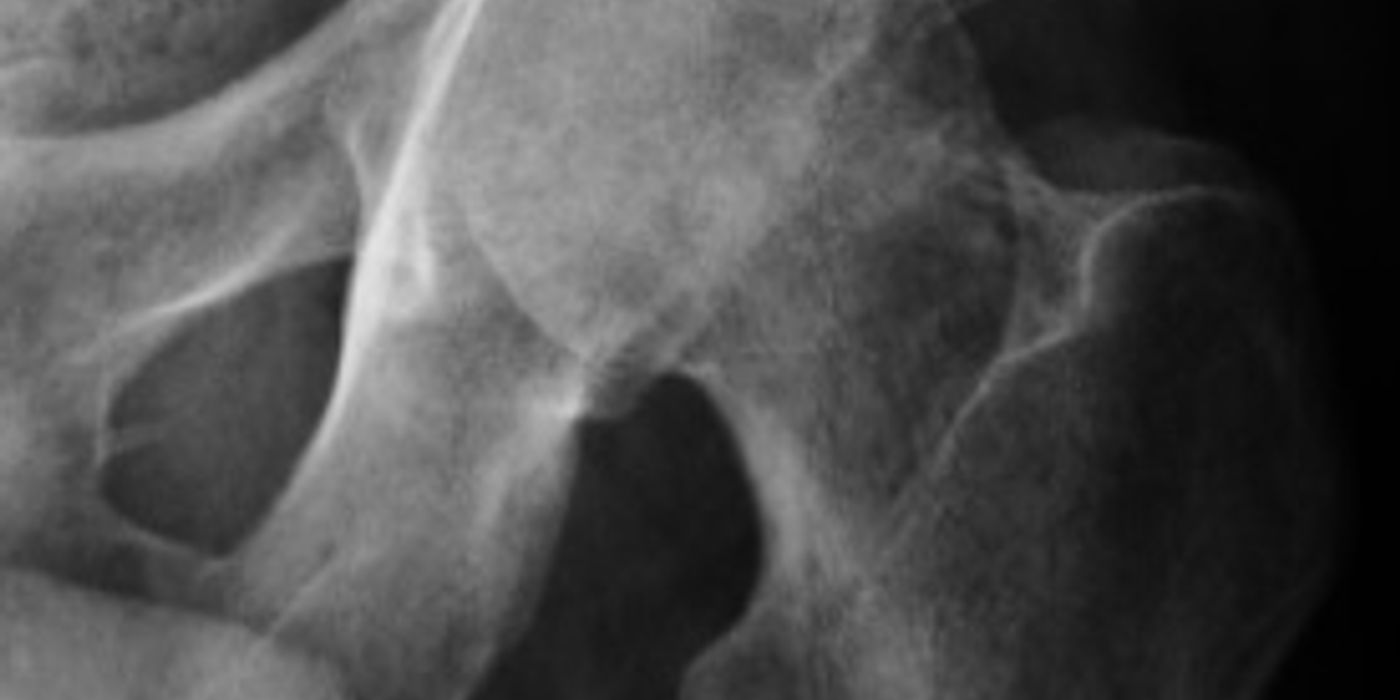
Septic arthritis is bacterial infection of a native (natural) synovial joint. It is an orthopaedic emergency: bacterial enzymes and the host inflammatory response destroy articular cartilage within days, and the reported case fatality in adults is around 11%. The core of management is to aspirate the joint before giving antibiotics (unless the patient is septic), then to perform urgent surgical drainage and washout with targeted antimicrobial therapy.
How does septic arthritis present, and who gets it?
The classic picture is a short history of a single hot, swollen, tender joint with severely restricted movement — often with fever, though its absence does not exclude infection. The 2006 BSR/BHPR/BOA/RCGP/BSAC guideline is explicit: such a patient "should be regarded as having septic arthritis until proven otherwise". The knee is the most commonly affected joint in adults; in children the hip predominates. Staphylococcus aureus is the most frequent organism, with streptococci next; Gram-negative organisms are seen in the elderly and immunosuppressed.
Risk factors and atypical hosts matter, both clinically and in interviews:
- Rheumatoid arthritis and other inflammatory arthropathies (a "flare" in a single joint should ring alarm bells)
- Diabetes mellitus, chronic kidney disease, immunosuppression (including biologics and steroids)
- Intravenous drug use — consider atypical joints (sacroiliac, sternoclavicular) and unusual organisms (including Gram-negatives and Candida)
- Extremes of age (over 80 and the very young)
- Recent joint surgery, arthroscopy or intra-articular injection; overlying skin breaks or ulcers
- Consider disseminated gonococcal infection in sexually active young adults, classically with migratory polyarthralgia, tenosynovitis and pustular skin lesions
How is septic arthritis diagnosed?
Diagnosis is clinical suspicion confirmed by joint aspiration. The 2006 BSR guideline mandates that synovial fluid "must be aspirated, Gram-stained and cultured prior to starting antibiotics", and the same principle is restated for the systemically well patient in the October 2023 BOAST on acute peri-prosthetic joint infection (which the July 2025 soft tissue infection BOAST applies to native joints): "A patient who is not septic should not be given antibiotics until appropriate deep tissue samples have been taken."
- Aspirate the joint under aseptic conditions and send fluid for urgent Gram stain, culture and sensitivities, white cell count, and polarising microscopy. The July 2025 BOAST states verbatim that the "aspirate should include samples for crystallography". Anticoagulation with warfarin is not a contraindication to needle aspiration (BSR 2006).
- Interpret the fluid cautiously. Frank pus or turbid fluid supports the diagnosis, and higher synovial white cell counts make sepsis progressively more likely (Margaretten et al., JAMA 2007). Crucially, per the BSR guideline, "neither the absence of organisms on Gram stain nor a negative subsequent synovial fluid culture excludes the diagnosis".
- Bloods: FBC, CRP, ESR, urea and electrolytes, liver function (to guide antibiotic dosing) and blood cultures before antibiotics. Serum urate has no diagnostic value in distinguishing acute gout from sepsis (BSR 2006).
- Imaging: plain radiographs as a baseline (they are usually normal early); ultrasound to confirm and guide aspiration of a deep effusion (hip); MRI if concomitant osteomyelitis or a periarticular collection is suspected. Imaging must never delay aspiration or drainage.
What else could it be?
The key differential is crystal arthropathy — gout and pseudogout can perfectly mimic sepsis, which is why crystallography is mandated. Remember that crystals and infection can coexist: identifying crystals does not exclude septic arthritis if suspicion remains. Other differentials include reactive arthritis, haemarthrosis, a monoarticular flare of rheumatoid or osteoarthritis, cellulitis or bursitis overlying a joint (do not aspirate through cellulitic skin), and Lyme arthritis. In children, consider transient synovitis, osteomyelitis, Perthes disease and SUFE.
How do children differ — and what are the Kocher criteria?
The commonest paediatric dilemma is the irritable hip: transient synovitis versus septic arthritis. Kocher and colleagues (JBJS Am, 1999) derived four independent predictors of hip sepsis in children: fever >38.5 °C, inability to weight-bear, ESR >40 mm/h and serum white cell count >12,000/mm³.
| Kocher predictors present | Predicted probability of septic arthritis (1999 derivation cohort) |
|---|---|
| 0 | <0.2% |
| 1 | 3% |
| 2 | 40% |
| 3 | 93.1% |
| 4 | 99.6% |
Caveats worth stating in an interview: the criteria were derived for the hip in one North American centre, and subsequent validation (including Kocher's own 2004 study) showed lower predictive performance — around 93% for four criteria. CRP has since emerged as one of the strongest independent predictors and is commonly added as a fifth criterion in modified versions. The criteria stratify risk; they do not replace aspiration when suspicion is real.
The May 2022 BOAST on children with acute musculoskeletal infection sets the UK standard: joint inpatient care by orthopaedic and paediatric teams; FBC, CRP, ESR and blood cultures with plain radiographs; ultrasound early and MRI as the preferred second-line modality "ideally within 48 hours"; empirical intravenous antibiotics started immediately in any child meeting high-risk sepsis criteria, but in the stable child listed for surgery "antibiotics may be delayed to allow deep tissue sampling". Confirmed septic arthritis in a child "requires surgical drainage as soon as possible, ideally within 24 hours of diagnosis", with clinical and radiographic follow-up for a minimum of 12 months to detect growth disturbance and avascular necrosis.
What is the initial management?
- Recognise sepsis first. As per the July 2025 BOAST, "a patient presenting with evidence of sepsis must have the 'sepsis six' protocol initiated immediately" (consistent with NICE NG51). In the septic patient, take blood cultures urgently and start broad-spectrum parenteral antibiotics without waiting for joint aspiration.
- In the systemically well patient, aspirate before any antibiotics, then start empirical intravenous therapy guided by the local microbiology policy (typically an anti-staphylococcal agent such as flucloxacillin, with cover modified for MRSA risk, Gram-negative risk, penicillin allergy or suspected gonococcus). Early microbiology involvement is a standard of care.
- Supportive care: analgesia, splint the joint in a position of comfort, keep the patient fasted for theatre, and ensure early senior orthopaedic review and consent for open as well as arthroscopic drainage. Document neurovascular status.
What is the definitive management?
The July 2025 BOAST states that "native joint infections should be treated according to standards outlined in the BOASt for the acute management of periprosthetic joint infection, recognising the time critical nature of chondral injury". Applying those standards:
- Urgent surgical drainage and lavage of the joint, performed by a suitably experienced surgical team as soon as it is safe — in the acutely unwell patient within 6 hours (Oct 2023 BOAST). Arthroscopic washout is standard for accessible joints such as the knee, shoulder and ankle; open washout remains appropriate where expertise or access dictates, and hip sepsis (particularly in children) usually requires urgent open drainage via an approach protecting the femoral head blood supply.
- Sample thoroughly in theatre: multiple deep tissue and fluid samples taken with separate sterile instruments and a no-touch technique before intra-operative antibiotics, mirroring the PJI BOAST sampling standard.
- Serial closed-needle aspiration to dryness is an accepted alternative in selected patients (BSR 2006) — for example gonococcal arthritis or patients unfit for surgery — but is not the default for a purulent large joint, and never for the hip.
- Antibiotic duration: conventionally up to two weeks intravenously followed by oral therapy for around four further weeks (BSR 2006), individualised with microbiology. The OVIVA trial (NEJM, 2019) showed oral antibiotics were non-inferior to intravenous therapy for complex bone and joint infection, and earlier oral switch under specialist guidance is now common practice. Repeat washout is indicated if the patient fails to improve clinically and biochemically.
- Rehabilitation: early physiotherapy and mobilisation once the joint is settling is good practice to limit stiffness.
What are the complications and prognosis?
- Mortality of approximately 11% in adults, higher in the elderly, polyarticular sepsis and delayed presentation (BSR 2006).
- Irreversible chondral loss leading to secondary osteoarthritis, chronic pain and stiffness; end-stage destruction may need arthrodesis or staged arthroplasty.
- Osteomyelitis, sinus formation and recurrent infection.
- In children: avascular necrosis of the femoral head, physeal arrest, limb length discrepancy and hip subluxation — hence the mandated 12-month minimum follow-up.
- Delay to drainage beyond 24–48 hours is consistently associated with worse joint outcomes, which is why every UK standard emphasises time-critical treatment.
Key points
- A hot, swollen, tender joint with restricted movement is septic arthritis until proven otherwise (BSR 2006).
- Aspirate before antibiotics in the well patient; in the septic patient, blood cultures then immediate antibiotics and the sepsis six.
- Always send aspirate for crystallography — crystal arthropathy is the great mimic, and crystals do not exclude coexisting infection.
- Kocher criteria (fever >38.5 °C, non-weight-bearing, ESR >40, WCC >12,000) stratify the paediatric irritable hip but do not replace aspiration.
- Native joints follow the acute PJI BOAST standards: urgent washout (within 6 hours if acutely unwell), multiple no-touch deep samples, and in children drainage ideally within 24 hours with 12 months' follow-up.
- Negative Gram stain or culture does not exclude the diagnosis; treat the patient, not the microbiology report.