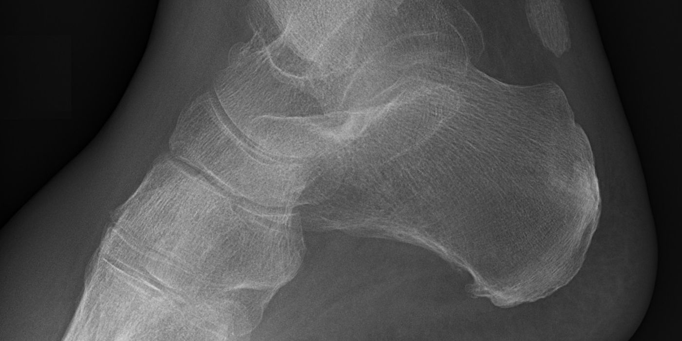
Hindfoot fractures are fractures of the calcaneus (heel bone) or talus, the two bones that connect the leg to the foot through the ankle and subtalar joints. They are typically high-energy injuries — a fall from height or a road traffic collision — and their outcome is determined as much by the soft-tissue envelope and joint surface damage as by the fracture itself. Displaced intra-articular calcaneal fractures are usually managed non-operatively in the UK following the Heel Fracture Trial (HeFT, BMJ 2014), whereas displaced talar neck fractures need urgent reduction and anatomical fixation because of the risk of avascular necrosis (AVN).
How do hindfoot fractures present and how are they diagnosed?
Both injuries follow axial loading: the calcaneus is crushed by a fall onto the heel; the talar neck fails under forced dorsiflexion against the anterior tibia (the classical "aviator's astragalus"). On assessment you should:
- Examine and document the soft tissues immediately — swelling, fracture blisters, skin tenting or threatened skin (especially over the posterior heel in tongue-type calcaneal fractures), and any open wound.
- Look for associated injuries of the same mechanism: lumbar spine fractures, contralateral hindfoot fractures, tibial plateau and pilon fractures. Around 10% of calcaneal fractures are bilateral and a similar proportion have spinal injury — image on suspicion.
- Document neurovascular status and assess repeatedly for foot compartment syndrome — pain out of proportion and pain on passive toe movement are the key early signs.
- Obtain radiographs (lateral and axial heel views; AP, lateral and Canale views for the talus). A reduced Böhler's angle (normally 20–40°) indicates posterior facet collapse.
- Obtain CT for any intra-articular fracture — it defines the posterior facet for Sanders classification of the calcaneus and improves the reliability of Hawkins grading of the talus, and it is essential for operative planning.
What is the initial management, and what do the soft tissues demand?
Initial management for both bones is elevation, ice, analgesia and a well-padded backslab, with serial soft-tissue review. Two BOA standards frame the emergencies:
- Open fractures — the BOA/BAPRAS open fracture BOAST (December 2017) explicitly covers open fractures of the "hind foot or midfoot" and requires intravenous prophylactic antibiotics "as soon as possible, ideally within 1 hour of injury", management in a centre providing combined orthoplastic care, debridement within 12 hours for solitary high-energy open fractures (immediately for gross contamination or vascular compromise), definitive soft-tissue cover within 72 hours if not achieved at first debridement, and internal fixation only when it can be immediately covered.
- Compartment syndrome — the July 2025 BOAST mandates hourly documented assessment of at-risk patients and immediate decompression once the diagnosis is made. For the foot specifically it states: "There is no consensus for the management of foot compartment syndrome. A documented justification of the management plan is required in these cases." In practice, decide and document: fasciotomy of the foot compartments, or a deliberate, justified decision to treat expectantly accepting possible claw-toe deformity.
For the talus, any fracture-dislocation (Hawkins II–IV) needs urgent reduction — in the emergency department under sedation where possible, or in theatre if irreducible — to decompress the soft tissues and the remaining blood supply. An extruded or button-holed talar body threatening the skin is a surgical emergency. This urgency is established good practice rather than a named guideline standard. NICE NG38 (Fractures — complex: assessment and management, 2016) aligns UK open-fracture care with the BOAST above.
How are calcaneal fractures classified and managed?
The Sanders classification (Sanders et al., 1993) grades displaced intra-articular fractures on the coronal CT cut through the widest part of the posterior facet:
| Sanders type | Posterior facet pattern |
|---|---|
| I | Non-displaced (<2 mm displacement), regardless of fragment number |
| II | Two-part (split) fracture |
| III | Three-part fracture with a central depressed fragment |
| IV | Four-part or highly comminuted articular fracture |
The pivotal UK evidence is the Heel Fracture Trial (HeFT) — Griffin et al., BMJ 2014: a pragmatic, assessor-blinded RCT of 151 adults with closed, displaced (≥2 mm posterior facet step) intra-articular calcaneal fractures across 22 UK centres, randomised to ORIF via an extensile lateral approach or non-operative care. At two years there was no difference in the primary outcome (Kerr-Atkins score 69.8 operative vs 65.7 non-operative; adjusted difference 0.0, 95% CI −7.1 to 7.0), and no difference in any secondary outcome including heel width, gait and return to work. Complications and reoperations were significantly more common with surgery (17/73, 23% vs 3/78, 4%; odds ratio 7.5, 95% CI 2.0 to 41.8), driven by a 19% surgical site infection rate. The authors concluded: "There is no justification to continue with this surgery for closed displaced intra-articular calcaneal fractures without severe displacement."
Practical management, therefore:
- Most closed, displaced intra-articular fractures: non-operative — elevation, early ankle and subtalar mobilisation as pain allows, six weeks non-weight-bearing then six weeks partial weight-bearing with physiotherapy (the HeFT protocol).
- Surgery is still indicated for the fractures HeFT excluded (good practice): open fractures, gross deformity with "fibula impingement" (severe varus/valgus with the fibular tip abutting or embedded in the calcaneus), tongue-type fragments threatening the posterior skin (urgent reduction to prevent necrosis), calcaneal fracture-dislocations, and displaced extra-articular tuberosity avulsions.
- Where fixation is undertaken, delay until the soft tissues settle (positive wrinkle test); percutaneous and sinus tarsi approaches reduce wound complications compared with the extensile lateral approach, though HeFT's caution about adopting them outside trials should be quoted.
How are talar neck fractures classified and managed?
The talar body is perfused retrogradely — dominantly by the posterior tibial artery via the artery of the tarsal canal and the deltoid artery, with contributions from the anterior tibial artery (head and neck) and the peroneal artery via the tarsal sinus — which is why progressive dislocation strips vascularity. The Hawkins classification (1970; Type IV added by Canale and Kelly, 1978) predicts AVN:
| Hawkins type | Pattern | AVN (Hawkins 1970) | AVN (pooled, Halvorson 2013) |
|---|---|---|---|
| I | Undisplaced neck fracture | 0% | 6% |
| II | Displaced, subtalar subluxation/dislocation | 42% | 18% |
| III | Subtalar and tibiotalar dislocation | 91% | 45% |
| IV | Subtalar, tibiotalar and talonavicular dislocation | — | 12% (rare; small numbers) |
Vallier et al. (JBJS Am, 2014) subdivided Type II: with subtalar subluxation only (IIA) no patients developed AVN, versus 25% after true subtalar dislocation (IIB) — supporting preservation of the deltoid artery as the key, and indicating that the initial displacement, rather than a modest delay to definitive fixation, drives osteonecrosis risk. Management:
- Hawkins I: CT to confirm no displacement; non-operative in cast, non-weight-bearing, or percutaneous screw fixation to allow earlier movement (good practice).
- Hawkins II–IV: urgent reduction of dislocations, then anatomical open reduction and stable fixation — typically dual anteromedial and anterolateral approaches to control neck alignment and avoid varus malunion, with plates and/or screws.
- Follow-up radiographs at 6–8 weeks for the Hawkins sign — subchondral lucency of the talar dome indicating preserved vascularity. It is highly sensitive for viability (100% in Tezval's series) but poorly specific (~58%), so its absence should prompt MRI rather than a presumption of AVN.
What are the complications and prognosis?
- Calcaneus: subtalar osteoarthritis, broadened heel with shoe-fitting problems, peroneal tendon impingement, and chronic pain; recovery plateaus at about 18 months and most HeFT patients returned to work (~85%), often to lighter duties. Wound breakdown and infection dominate operative complications. Late subtalar fusion salvages painful arthritis (performed in three non-operative HeFT patients by two years).
- Talus: AVN with dome collapse (31% in Vallier's 100-fracture series), post-traumatic ankle (18%) and subtalar (15%) arthritis, varus malunion from medial comminution, and non-union. Established collapse may need hindfoot fusion.
Key points
- Hindfoot fractures are soft-tissue injuries with a bone problem: apply the open fracture BOAST (antibiotics within 1 hour, orthoplastic care) and the July 2025 compartment syndrome BOAST, which requires a documented justification for the chosen management of foot compartment syndrome.
- HeFT (BMJ 2014): ORIF of typical closed, displaced intra-articular calcaneal fractures gives no functional benefit at two years and seven-fold higher odds of complications — non-operative care is the UK default.
- Surgery retains a role in calcaneal fractures HeFT excluded: open injuries, fracture-dislocations, fibular impingement and skin-threatening tongue-type fragments.
- Sanders (CT) classifies calcaneal fractures; Hawkins (I–IV) predicts talar AVN — roughly 0%, 20–40%, 45–90% for types I–III.
- Reduce talar fracture-dislocations urgently; fix talar neck fractures anatomically, often via dual approaches; check for the Hawkins sign at 6–8 weeks.