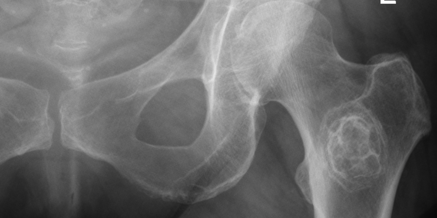
Metastatic bone disease (MBD) is the spread of a primary cancer — most commonly breast, prostate, lung, renal or thyroid — to the skeleton, where it causes pain, hypercalcaemia, pathological or impending fracture, and spinal cord compression. In the UK its management is defined by the June 2022 BOAST Management of Metastatic Bone Disease, which mandates full investigation of any lesion of unknown primary before fixation, multidisciplinary decision-making, and surgical constructs that allow immediate weight-bearing and outlast the patient. Fracture risk in long-bone lesions is estimated with the Mirels score, with prophylactic fixation generally recommended at a score of 9 or more.
When should you suspect metastatic bone disease?
The Jun 2022 BOAST lists the red flags that should trigger suspicion of malignancy in a presenting fracture or painful bone: "low energy fractures in the non-osteoporotic population, antecedent pain, night pain, absence of injury, and insidious pain". Standard 3 requires that "prodromal pain, history of malignancy, or night pain raise suspicion of MBD and should be documented along with any circumstances of injury".
The commonest primaries are breast, prostate, lung, kidney and thyroid (with myeloma the key haematological mimic to exclude), and the commonest skeletal sites are the spine, pelvis, and proximal femur. The BOAST covers the appendicular skeleton, pelvis and scapula; osteoporotic fractures and spinal metastases are outside its scope, the latter following the NICE pathway below.
How is a bone lesion of unknown primary investigated before fixation?
This is the single most examined principle: never fix a solitary bone lesion until a primary bone tumour has been excluded. Nailing an unrecognised sarcoma contaminates the whole bone and soft-tissue compartments and can convert a limb-salvageable tumour into one requiring amputation. The BOAST work-up when MBD is suspected (standard 6) is, verbatim:
- "FBC, U+E, LFT, calcium & bone profile, PSA in men, myeloma screen"
- "Orthogonal radiographs of the whole bone"
- "Staging CT of the thorax, abdomen and pelvis (CT-TAP) within 24 hours of orthopaedic assessment"
The pathway then branches on the findings:
- Radiographic features of a primary bone tumour (bone destruction, new bone formation, periosteal reaction, soft-tissue swelling) → referral to a bone sarcoma centre within 72 hours (standard 4).
- A CT-TAP without evidence of malignancy may itself indicate a primary bone tumour and also requires sarcoma-centre referral within 72 hours (standard 7).
- "Biopsy of a suspected primary bone tumour must be performed at a bone sarcoma centre" (standard 5) — never in the district general hospital, because the biopsy track must be planned so it can be excised at definitive surgery.
- MBD without an obvious primary should be discussed with the local acute oncology service (standard 8), and a solitary bone metastasis requires referral to a recognised tertiary centre (standard 9) — a solitary renal or thyroid metastasis may be better treated by en-bloc resection and endoprosthetic replacement than by intralesional fixation.
A patient with a known disseminated malignancy and multiple typical metastases does not need biopsy of every lesion, but the blood panel and staging CT still inform prognosis, anaesthetic risk and oncological options — and multidisciplinary decisions on (neo)adjuvant therapy should be recorded before surgery (standard 10).
What is the Mirels score and when is prophylactic fixation indicated?
Mirels (Clin Orthop Relat Res, 1989) proposed a weighted score, 4–12, to predict pathological fracture in long-bone metastases and guide prophylactic fixation before radiotherapy. Four variables each score 1–3:
| Variable | 1 point | 2 points | 3 points |
|---|---|---|---|
| Site | Upper limb | Lower limb | Peritrochanteric |
| Pain | Mild | Moderate | Functional (mechanical, on weight-bearing) |
| Lesion type | Blastic | Mixed | Lytic |
| Size (fraction of bone diameter) | < 1/3 | 1/3 – 2/3 | > 2/3 |
Interpretation: a score of ≤ 7 carries a low fracture risk and can be managed with radiotherapy and observation; a score of 8 is borderline (about 15% fracture risk) and warrants case-by-case MDT discussion; a score of ≥ 9 (33% fracture risk at 9 in the original series) is an impending fracture and prophylactic fixation is recommended before irradiation. It is good practice to add clinical judgement — functional pain despite radiotherapy, and lytic lesions in the peritrochanteric femur, are treated with a low threshold for fixation, and prophylactic surgery is generally associated with easier surgery and better outcomes than fixing a completed pathological fracture.
What are the principles of definitive surgical management?
Two BOAST standards carry most of the weight and are worth quoting verbatim in an interview. Standard 11: "Surgery for MBD should be consultant led." Standard 12: "Surgical interventions should outlast the lifetime of the patient. Where internal fixation is used, curettage and cement augmentation is recommended to replace bone loss. All patients require a construct to allow immediate weight-bearing."
In practice this means:
- Protect the whole bone — long intramedullary nails spanning the femur or humerus rather than short constructs, because further metastases may develop distally; this is established good practice consistent with the "outlast the patient" standard.
- Arthroplasty over fixation where bone stock is destroyed — cemented hemiarthroplasty or total hip replacement (often with a long stem) for femoral neck and head lesions, and endoprosthetic replacement for massive peritrochanteric destruction or solitary resectable metastases.
- Curettage and cement augmentation of the defect when internal fixation is used, per standard 12.
- Thromboprophylaxis for all: "All patients require thromboprophylaxis. Contraindications must be documented" (standard 13) — malignancy plus lower-limb surgery is a very high VTE-risk combination.
- Post-operative radiotherapy to the operated segment is standard oncological practice to control local progression, decided with the MDT (standards 10 and 16).
How is metastatic spinal cord compression managed?
Spinal metastases sit outside the BOAST and follow NICE guideline NG234, Spinal metastases and metastatic spinal cord compression (2023, replacing CG75). The pathway for suspected MSCC — back pain with neurological symptoms or signs in a patient with cancer — is an oncological emergency:
- Contact the MSCC coordinator the same day and treat as an emergency; immobilise (flat bed rest with spinal precautions) if symptoms or signs suggest spinal instability.
- Give 16 mg oral dexamethasone (or equivalent parenteral dose) as soon as possible, continued daily while awaiting definitive treatment, with gastric protection and glucose monitoring.
- Perform urgent whole-spine MRI, ideally within 24 hours of suspicion.
- Deliver definitive treatment — surgery or radiotherapy — urgently, ideally before further neurological deterioration; ambulatory status at treatment is the strongest predictor of walking afterwards.
The landmark evidence for surgery is Patchell et al. (Lancet, 2005): 101 patients randomised to direct decompressive surgery plus radiotherapy versus radiotherapy alone; 84% versus 57% could walk after treatment, and walkers retained ambulation for a median of 122 versus 13 days. Surgical candidacy depends on prognosis, fitness and a single level of compression, decided jointly with spinal surgery and oncology.
What are the complications, and how are patients followed up?
Beyond fixation failure and the usual perioperative risks, key complications are VTE, hypercalcaemia (confusion, polyuria, constipation — treat with fluids and bisphosphonates), marrow infiltration and anaemia, and disease progression around the implant. The BOAST closes the loop on follow-up: patients with ongoing pain "should continue under orthopaedic surveillance" as this may indicate "disease progression and/or impending failure of the reconstruction" (standard 14); "failed MBD surgical intervention must be discussed with a recognised tertiary centre" (standard 15); and decisions on adjuvant therapy, rehabilitation and palliation "should involve the patient, their family, and carers" (standard 16). Every unit needs an agreed MDT policy and network referral pathways (standards 1–2).
Key points
- Red flags: low-energy fracture in a non-osteoporotic patient, antecedent, night or insidious pain, absence of injury.
- Never fix a lesion of unknown primary: bloods (including PSA and myeloma screen), orthogonal whole-bone radiographs and CT-TAP within 24 hours first; suspected primary bone tumours go to a sarcoma centre within 72 hours and are biopsied only there.
- Solitary metastases and MBD with no obvious primary need tertiary/acute-oncology referral before surgery.
- Mirels score ≥ 9 = impending fracture → prophylactic fixation before radiotherapy; 8 is borderline; ≤ 7 favours radiotherapy and observation.
- Surgery is consultant-led; constructs must outlast the patient, allow immediate weight-bearing, and use cement augmentation with internal fixation; all patients get thromboprophylaxis.
- Suspected MSCC: 16 mg dexamethasone immediately, whole-spine MRI within 24 hours, MSCC coordinator, and urgent definitive surgery or radiotherapy (Patchell 2005 supports decompression plus radiotherapy in selected patients).