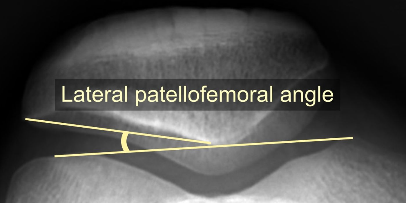
A first-time (lateral) patellar dislocation is an acute injury in which the patella displaces completely out of the trochlear groove, almost always laterally, tearing the medial patellofemoral ligament (MPFL) and frequently causing a haemarthrosis. It is managed with prompt reduction, radiographs including a skyline view, early mobilisation in a flexion-permitting splint rather than a cast, and structured physiotherapy; surgery is reserved for associated injuries such as osteochondral fractures, not for routine stabilisation. UK practice is defined by the British Orthopaedic Association's BOAST Assessment and Management of First Time Lateral Patellar Dislocation (FTLPD), published December 2024 and endorsed by BASK and BSCOS.
Who gets it, and what actually tears?
First-time lateral patellar dislocation is predominantly an injury of adolescents and young adults, typically occurring during sport through a twisting mechanism on a flexed, valgus knee or, less often, a direct blow. The essential lesion is failure of the MPFL, the primary passive restraint to lateral patellar translation, often accompanied by an osteochondral or chondral injury as the patella relocates across the lateral femoral condyle. Underlying anatomical variants — trochlear dysplasia, patella alta, an increased tibial tuberosity–trochlear groove (TT-TG) distance and generalised ligamentous laxity — predispose to both the first event and to recurrence.
How does it present, and how is it diagnosed?
Many patellae relocate spontaneously or with knee extension before arrival, so the classic presentation is a painful, swollen knee after a twisting injury with a tense effusion (haemarthrosis) rather than an obviously deformed joint. Assess for:
- Medial retinacular tenderness (over the torn MPFL) and a positive patellar apprehension test once acute pain settles.
- A large haemarthrosis — in a young patient this should prompt consideration of patellar dislocation alongside ACL rupture and osteochondral fracture.
- Recurrence risk factors, which the BOAST requires to be documented: demographics, mechanism of injury, any history of contralateral dislocation, hypermobility and family history, plus examination of generalised joint laxity and lower limb alignment.
After reduction, the imaging standard is an AP and lateral radiograph of the knee, including a skyline patellar view. The skyline view is specifically mandated because it is the projection most likely to reveal medial patellar facet or lateral femoral condyle osteochondral fragments and residual subluxation; a loose body may be the only radiographic clue to a significant chondral injury.
What is the initial management?
The December 2024 BOAST sets out a deliberately conservative, motion-preserving pathway:
- Reduce promptly. "An acute lateral patellar dislocation should be reduced without delay." Reduction is usually achieved with analgesia, progressive knee extension and gentle medially directed pressure on the patella.
- Do not aspirate. "Aspiration of an associated haemarthrosis should not be performed." Aspiration adds infection risk without changing management (good-practice rationale; the standard itself is unqualified).
- Image after reduction with AP, lateral and skyline views as above.
- No cast. "Cast immobilisation should not be used, but splints that permit full knee flexion may be used for pain relief in the period immediately following injury." Rigid immobilisation promotes stiffness and quadriceps inhibition without reducing recurrence.
- Mobilise immediately. "Unrestricted weight bearing should be encouraged immediately, with the aid of crutches if required."
- Early review. Clinical evaluation by an appropriately qualified professional within 2 weeks, in an age-appropriate setting (children should be seen in a paediatric-appropriate service), with the recurrence risk factors above documented.
This is a genuine change in emphasis from historical practice, where cylinder casts and aspiration of tense effusions were common: the standard explicitly rules both out.
When is an MRI indicated?
MRI is not routine for every first-time dislocation. As per the Dec 2024 BOAST, MRI (when not contraindicated) should be obtained when:
- an associated injury (such as an osteochondral fracture or loose body) is suspected;
- alternative diagnoses cannot be excluded clinically;
- the patient is difficult to assess;
- surgery is being planned; or
- conservative treatment has failed.
What is the definitive management?
Rehabilitation first
The mainstay of treatment for an isolated first-time dislocation is non-operative: assessment by a musculoskeletal physiotherapist within 3 weeks of injury, followed by a progressive programme restoring range of movement, quadriceps (particularly vastus medialis obliquus) strength, and proximal hip and core control before graded return to sport. Patient education resources should be available as part of the pathway.
Surgery — for associated injury, not routine stabilisation
- Surgery for associated injuries, classically a displaced osteochondral fracture or intra-articular loose body, may be considered — typically fragment fixation where the fragment is salvageable, or removal where it is not (the choice of technique is good practice rather than BOAST-specified).
- "Surgery solely to stabilise the patella should not be offered routinely following isolated FTLPD." First-time MPFL reconstruction is therefore not standard UK practice outside these indications.
- "Isolated lateral retinacular release is never indicated." This is one of the few absolute prohibitions in any BOAST and a frequent interview discussion point.
- Where surgery is being considered, the case requires multidisciplinary team review — particularly relevant for osteochondral fragments in skeletally immature patients, where decisions sit between paediatric knee surgeons, physiotherapists and, where appropriate, tertiary patellofemoral services.
Follow-up
Patient-initiated follow-up is acceptable for most patients. The exceptions requiring planned review are those undergoing surgery, those failing conservative treatment, those at high risk of recurrence, and children with significant lower limb malalignment.
What is the risk of recurrence, and who is at risk?
Recurrence is the key long-term complication. In a systematic review and meta-analysis of 17 studies (Huntington et al., American Journal of Sports Medicine 2020), the overall recurrence rate after a first-time lateral patellar dislocation was 33.6%, with risk strongly stratified by anatomical and demographic factors:
| Risk factor | Odds ratio for recurrence |
|---|---|
| Trochlear dysplasia | 4.15 |
| Elevated TT-TG distance | 2.87 |
| Open physes | 2.72 |
| Younger age | 2.61 |
| Patella alta | 2.38 |
Risk is cumulative: recurrence was roughly 8–14% with no risk factors, 30–60% with two, and 70–79% with three. Notably, sex, the pattern of MPFL injury and a history of contralateral dislocation were not significantly associated with recurrence in the pooled analysis. Patients who go on to recurrent instability move onto a different pathway, covered by the separate BOA standards on the assessment and surgical management of recurrent patellar instability (August 2020), where MPFL reconstruction and bony realignment procedures have their place. Other recognised sequelae of the first event include osteochondral damage, persistent anterior knee pain and long-term patellofemoral osteoarthritis.
Why does this matter for the ST3 interview?
This is a current, guideline-anchored topic that tests whether a candidate manages a "benign" injury to a modern standard: the discriminating answers are no aspiration, no cast, skyline view, physiotherapy within 3 weeks, MRI only for defined indications, MDT review before any surgery, and the two prohibitions — no routine first-time stabilisation and never an isolated lateral release — each anchored explicitly to the December 2024 BOAST.
Key points
- Reduce an acute lateral patellar dislocation without delay; do not aspirate the haemarthrosis.
- Post-reduction imaging: AP and lateral radiographs including a skyline patellar view.
- No cast — use a splint permitting full knee flexion for pain relief only, with immediate unrestricted weight bearing.
- Clinical review within 2 weeks (age-appropriate setting) and physiotherapy assessment within 3 weeks.
- MRI for suspected associated injury, diagnostic uncertainty, surgical planning or failed conservative treatment — not routinely.
- Surgery is for associated injuries such as osteochondral fractures, after MDT review; stabilisation surgery is not offered routinely for an isolated first dislocation, and isolated lateral release is never indicated.
- Overall recurrence is about one in three; trochlear dysplasia, elevated TT-TG, open physes, younger age and patella alta multiply the risk.