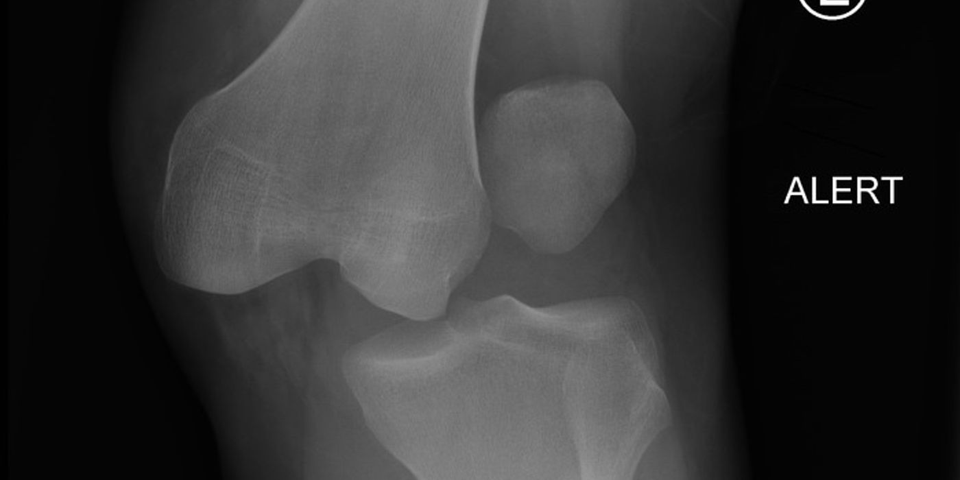
A knee dislocation is complete loss of the tibiofemoral articulation and, by definition, a multiligament knee injury: it requires rupture of at least two of the four major ligament complexes (ACL, PCL, MCL, LCL/posterolateral corner). It is a limb-threatening orthopaedic emergency because the tethered popliteal artery is injured in roughly 5–21% of cases, and management centres on urgent reduction, rigorous vascular assessment and revascularisation within one hour of arrival where the artery is injured, followed by staged or early ligament reconstruction.
How does a knee dislocation present, and why is it easily missed?
Around half of knee dislocations reduce spontaneously before arrival, so a normally aligned knee does not exclude the diagnosis. Suspect a reduced dislocation in any knee with gross multidirectional laxity, a large haemarthrosis after high-energy trauma, or a low-energy twisting injury in a patient with obesity — an increasingly recognised UK cohort. Common peroneal nerve injury accompanies 15.9–25% of multiligament knee injuries, so a documented neurological examination is essential at presentation and after every manipulation; persistent or evolving deficits should be managed along the pathway set out in the BOAST on peripheral nerve injury (December 2021).
Examine for the dimple sign — skin puckering over the medial joint line in a posterolateral dislocation, indicating buttonholing of the medial femoral condyle through the capsule. This pattern is frequently irreducible closed and risks skin necrosis; it is an indication for urgent open reduction (good practice, not a guideline standard).
How is a knee dislocation classified?
The Schenck (Kennedy–Schenck) anatomical classification grades the injury by the ligaments torn, with higher grades reflecting greater energy and worse prognosis. Vascular (C) and neurological (N) modifiers are appended (e.g. KD IIIL-C).
| Grade | Pattern |
|---|---|
| KD I | Multiligament injury with one cruciate intact (ACL or PCL spared) |
| KD II | Both cruciates ruptured, collaterals intact |
| KD III | Both cruciates plus one collateral complex — IIIM (medial) or IIIL (lateral/posterolateral corner) |
| KD IV | All four ligament complexes ruptured |
| KD V | Multiligament injury with periarticular fracture (fracture-dislocation) |
How should the vascular status be assessed?
Vascular assessment is governed by the BOAST on diagnosis and management of arterial injuries associated with musculoskeletal trauma (June 2026), which states that "clinical arterial examination is mandatory and specific findings and timings must be documented" and that "a pulseless deformed limb must be urgently realigned and splinted. Arterial examination must be repeated and documented." Where arterial injury is suspected, consultant input is mandatory.
Hard signs of arterial injury mandate immediate senior vascular involvement rather than further imaging-driven delay:
- Absent or diminished distal pulses after reduction
- Active haemorrhage or an expanding/pulsatile haematoma
- An audible bruit or palpable thrill
- Distal ischaemia — pain, pallor, paraesthesia, paralysis, coldness
In the pulse-present knee, measure the ankle–brachial pressure index (ABPI). In Mills and colleagues' prospective series (Journal of Trauma, 2004), an ABPI <0.90 identified every surgically significant arterial injury, and an ABPI ≥0.90 had a 100% negative predictive value when combined with serial examination. An ABPI threshold of 0.90 is therefore established good practice, though not itself a numbered BOAST standard: below 0.90, obtain urgent arterial imaging; at or above 0.90, admit for serial vascular observations for 24–48 hours. The 2026 BOAST directs the imaging choice: "CT angiogram is recommended if arterial injury is suspected and should be performed concurrently with whole-body CT" — imaging must never delay revascularisation of the ischaemic limb.
What is the initial management of a dislocated knee?
- Reduce urgently under sedation or anaesthesia, splint in 20–30° of flexion, and repeat and document the vascular and neurological examinations with timings.
- Control haemorrhage by direct pressure, packing or a distally applied tourniquet; the BOAST prohibits blind clamping.
- Revascularise within one hour where the artery is injured: "Revascularisation is an emergency procedure (NCEPOD 1) and should be commenced within one hour of arrival to hospital." Consultant-led care from orthopaedics together with vascular or plastic surgery is mandatory in confirmed injury.
- Shunt, fix, then repair. The BOAST recommends a temporary intravascular shunt to restore flow rapidly, then "rapid skeletal reduction and stabilisation should be achieved immediately following reperfusion and prior to definitive revascularisation" — in practice a knee-spanning external fixator — followed by arterial repair or an interposition (usually reversed saphenous) graft. Bypass grafts are reserved for blast or ballistic injury or where anatomical reconstruction is not feasible.
- Fasciotomy: "Fasciotomies must always be considered after revascularisation. Any contrary decision should be documented with the name of the senior decision maker." Thereafter maintain vigilance for compartment syndrome as per the July 2025 BOAST, with hourly documented assessment of the at-risk limb and immediate decompression on diagnosis.
- Spanning external fixation is also indicated without vascular injury for open dislocation, gross instability that cannot be held reduced in a splint, and in patients whose body habitus defeats bracing (good practice).
- Where cognition allows, patients must be made aware of the possibility of amputation; any early amputation decision requires two consultants and clear documentation.
How is the multiligament injury managed definitively?
Once the limb is safe, obtain MRI to map the ligamentous injury, and stress radiographs or examination under anaesthesia to grade instability. Definitive management is not covered by a BOAST; the following reflects contemporary consensus and good practice:
- Operative treatment is preferred in active patients — modern series consistently show better function than bracing alone.
- Reconstruction is generally favoured over isolated repair for the cruciates and the posterolateral corner, where repair alone has historically higher failure rates; avulsions with good tissue may be repaired and augmented.
- Timing: early single-stage surgery (within roughly three weeks) suits most patterns; staged surgery (early lateral/posterolateral work, delayed cruciate reconstruction) is used where swelling, skin or capsular healing dictate. Vascular repair usually delays ligament surgery until the graft is secure.
- Rehabilitation balances graft protection against the dominant complication, arthrofibrosis: early protected range of movement in a hinged brace, with quadriceps activation from the outset.
How does isolated ACL rupture differ?
An isolated ACL rupture is a sports injury, not a limb-threatening one, presenting with a pop, immediate haemarthrosis and rotatory instability. Management is either structured rehabilitation or arthroscopic reconstruction (hamstring, patellar tendon or quadriceps autograft). The UK ACL SNNAP trial (Lancet, 2022) randomised 316 patients with non-acute ACL injury and persistent instability: surgical reconstruction was clinically superior to rehabilitation-first management (mean KOOS4 73.0 vs 64.6 at 18 months), and 41% of the rehabilitation arm crossed over to surgery. Rehabilitation remains reasonable first-line for lower-demand patients without ongoing instability.
What are the complications and prognosis?
- Stiffness/arthrofibrosis — the most common complication after multiligament reconstruction.
- Common peroneal nerve palsy — frequently incomplete recovery; manage per the peripheral nerve injury BOAST.
- Vascular compromise, compartment syndrome and amputation — amputation complicates around 2.5% of multiligament knee injuries, concentrated in delayed or missed arterial injury.
- Residual laxity, venous thromboembolism and post-traumatic osteoarthritis in the longer term. Return to pre-injury sport is the exception rather than the rule after high-grade injuries.
Key points
- A knee dislocation is a multiligament injury; half arrive reduced — suspect it in any grossly unstable knee.
- Documented, timed arterial examination is mandatory; re-examine after every reduction (Jun 2026 BOAST).
- ABPI <0.90 → urgent CT angiography; ≥0.90 → serial examination (Mills 2004; CTA per the 2026 BOAST).
- Revascularisation is NCEPOD 1: start within one hour of arrival — shunt, spanning external fixation, definitive repair or graft, and always consider fasciotomy.
- Classify with Schenck KD I–V; reconstruct rather than brace in active patients, watching for arthrofibrosis.
- For isolated non-acute ACL rupture with instability, ACL SNNAP supports reconstruction over rehabilitation-first management.