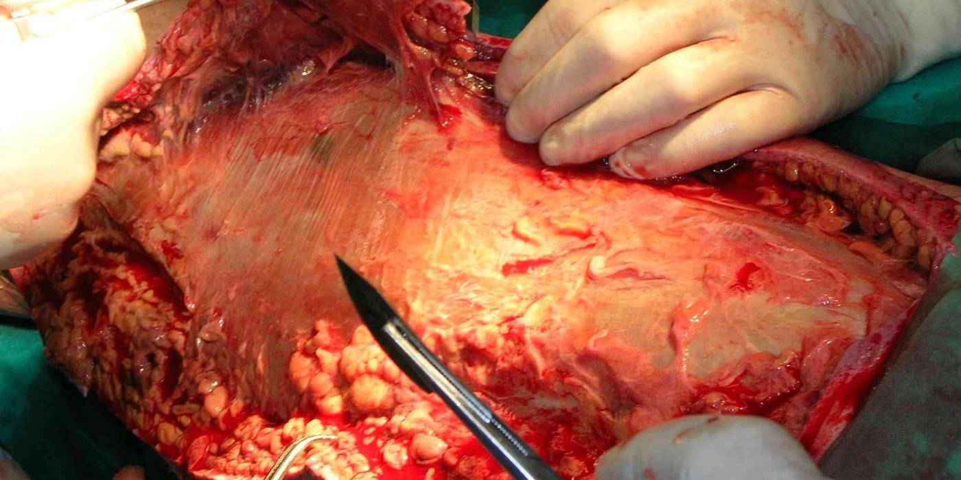
Necrotising fasciitis is a rapidly progressive, life-threatening bacterial infection that spreads along the deep fascial planes, causing necrosis of fascia and overlying soft tissue with severe systemic sepsis. It is a clinical diagnosis and a surgical emergency: no blood test, score or scan reliably excludes it, and survival depends on immediate radical surgical debridement combined with broad-spectrum antibiotics and aggressive sepsis care. UK management is defined by the BOA/BAPRAS standard Management of Musculoskeletal Soft Tissue Infections (BOAST, July 2025), which states that surgery "is required urgently and should not be delayed by medical and intensive care management, imaging, or inter-hospital transfer".
How does necrotising fasciitis present, and how is it diagnosed?
The cardinal early feature is pain out of proportion to the visible skin changes, often with rapid progression over hours. Early appearances mimic cellulitis; the discriminating features are severity of pain, tenderness extending beyond the erythema, systemic toxicity disproportionate to the local signs, and failure to respond to antibiotics. Late ("hard") signs — haemorrhagic bullae, fixed skin staining, crepitus, anaesthesia of the skin and frank necrosis — indicate established disease.
Two broad microbiological patterns are recognised:
- Type 1 (polymicrobial) — mixed aerobes and anaerobes, typically in patients with diabetes, immunosuppression or chronic disease, and more common on the trunk and perineum (Fournier's gangrene is the perineal variant).
- Type 2 (monomicrobial) — classically group A Streptococcus (sometimes with Staphylococcus aureus), affecting previously healthy patients of any age, usually in a limb, and the pattern most often seen in orthopaedic practice.
English Hospital Episode Statistics data show the condition is rare but increasing: age-standardised incidence roughly doubled from 9 to 21 per million between 2002 and 2017, with in-hospital mortality static at around 16% (Bodansky et al., 2020).
What is the role of the LRINEC score, and what are its limitations?
The Laboratory Risk Indicator for Necrotising Fasciitis (LRINEC; Wong et al., 2004) is a 13-point score derived from six routine tests — CRP, white cell count, haemoglobin, sodium, creatinine and glucose — with a score of 6 or more suggesting necrotising infection. The derivation study reported a sensitivity of 89.9% and specificity of 96.9%, but subsequent validation studies have reported sensitivities as low as 36–83%, so a low LRINEC score cannot rule out necrotising fasciitis. The July 2025 BOAST deliberately makes no mention of scoring systems: diagnosis is clinical, and where doubt remains the definitive investigation is surgical exploration of the fascia, not further imaging or bloods. Imaging (gas on plain films or CT) is specific when present but insensitive, and waiting for it delays surgery. In interview and in practice, LRINEC should be described as an adjunct that may raise suspicion, never a gatekeeper to theatre.
What is the immediate management?
Initial management runs on two parallel tracks — resuscitation and mobilisation for surgery — and the BOAST requires every hospital to have a locally agreed multi-specialty pathway that names the specialty with primary clinical responsibility, so ownership is never ambiguous.
- Sepsis care: per the BOAST, "a patient presenting with evidence of sepsis must have the 'sepsis six' protocol initiated immediately" — oxygen, blood cultures, IV broad-spectrum antibiotics, IV fluids, lactate and urine output monitoring — consistent with NICE guideline NG51 (Suspected sepsis: recognition, diagnosis and early management).
- Antibiotics: broad-spectrum parenteral antibiotics started immediately, guided by the local microbiologist. Antibiotics alone will not control necrotising infection because the thrombosed microcirculation cannot deliver them to dead tissue; they are an adjunct to surgery, not an alternative.
- Immediate surgery: the BOAST is explicit that surgery "should not be delayed by medical and intensive care management, imaging, or inter-hospital transfer". The patient should go to theatre at the admitting hospital, out of hours if necessary, operated on by "suitably qualified surgeon(s)". Good practice is senior joint operating (orthopaedics with plastics or general surgery according to site) rather than delegation or deferral.
- Patients unfit for surgery: where surgery is not in the patient's interest, the BOAST requires a documented rationale and referral for end-of-life care — an active, senior decision rather than a default.
What does definitive surgical management involve?
The operation is a radical debridement of all necrotic and infected tissue back to healthy, bleeding, viable margins. The BOAST requires a systematic, documented assessment of the anatomical structures involved, and its advisory notes describe three tissue zones to guide the extent of excision:
| Zone | BOAST description | Surgical action |
|---|---|---|
| Zone 1 | "The area of obvious necrosis, with fixed staining, thrombosed blood vessels or haemorrhagic bullae" | Excise completely |
| Zone 2 | "Areas of likely infection with warm, red skin, exquisite tenderness, or induration" | Explore and debride until healthy tissue is reached |
| Zone 3 | "Normal skin and fascia" | Preserve; defines the margin of resection |
Key intra-operative and early post-operative standards from the July 2025 BOAST:
- Deep tissue sampling: multiple deep tissue samples must be taken and labelled as "necrotising infection" so the laboratory prioritises and processes them appropriately.
- Amputation decisions: where life-saving amputation is considered, the decision requires at least two consultants from different specialties.
- No primary closure: "primary closure should not be performed at the index operation" — wounds are left open with appropriate dressings, anticipating a planned return to theatre.
- Early review: the treating team must review the patient within 6 hours of diagnosis or surgery.
- Mandatory re-exploration if not improving: "failure to improve after surgical debridement implies incomplete resection of infected tissue and should mandate further surgical exploration." A planned second look at 24–48 hours is standard practice, with repeated debridements until the wound is clean and the patient is improving.
Postoperatively the patient is managed in a critical care setting with ongoing organ support, microbiology-directed rationalisation of antibiotics once cultures return, and correction of coagulopathy, anaemia and nutrition. In group A streptococcal disease, clindamycin is commonly added for its toxin-suppressing effect, and intravenous immunoglobulin may be considered in streptococcal toxic shock — both are microbiology-led decisions rather than BOAST standards.
How are the resulting defects reconstructed?
Radical debridement produces large soft-tissue defects, so reconstruction should be planned from the outset with early plastic surgical involvement (good practice, in keeping with the standard's BOA/BAPRAS co-authorship). Once the infection is controlled and the wound bed is clean, the reconstructive ladder applies: negative-pressure wound therapy as a bridge, delayed direct closure, split-skin grafting for most fascial defects, and local or free flap cover where tendon, bone or neurovascular structures are exposed. Amputees and patients with major functional loss need early rehabilitation input, and all survivors need honest counselling — scarring, contracture and psychological morbidity are the norm rather than the exception.
What are the complications and prognosis?
- Mortality in English cohort data is around 16% overall, and considerably higher with established septic shock or delayed surgery; historical series report 30% or more.
- Morbidity includes amputation, multi-organ failure, prolonged critical care stay, large scarred defects, contractures and long-term psychological sequelae.
- The dominant modifiable prognostic factor is time to first adequate debridement — the entire structure of the July 2025 BOAST exists to compress that interval.
The same BOAST also sets standards for the wider spectrum of severe soft-tissue infection: native large joint infections should be managed in line with the peri-prosthetic joint infection BOAST (October 2023); limb cellulitis is managed by a single named specialty on a locally agreed pathway; simple abscesses in well patients may be drained under local anaesthesia, while deep or complex abscesses require formal drainage in an operating theatre.
Key points
- Necrotising fasciitis is a clinical diagnosis — pain out of proportion, rapid progression and systemic toxicity; hard signs are late.
- LRINEC and imaging cannot exclude the diagnosis and must never delay surgery; if in doubt, explore the fascia.
- Per the July 2025 BOAST: start the sepsis six and broad-spectrum IV antibiotics immediately, and operate urgently without delay for imaging, ICU optimisation or transfer.
- Debride radically using the three-zone assessment, take multiple deep samples labelled "necrotising infection", and do not close primarily.
- Review within 6 hours; failure to improve mandates further surgical exploration; amputation decisions need two consultants from different specialties.
- Reconstruction is planned early with plastic surgery; mortality is around 16% in England and hinges on time to adequate debridement.