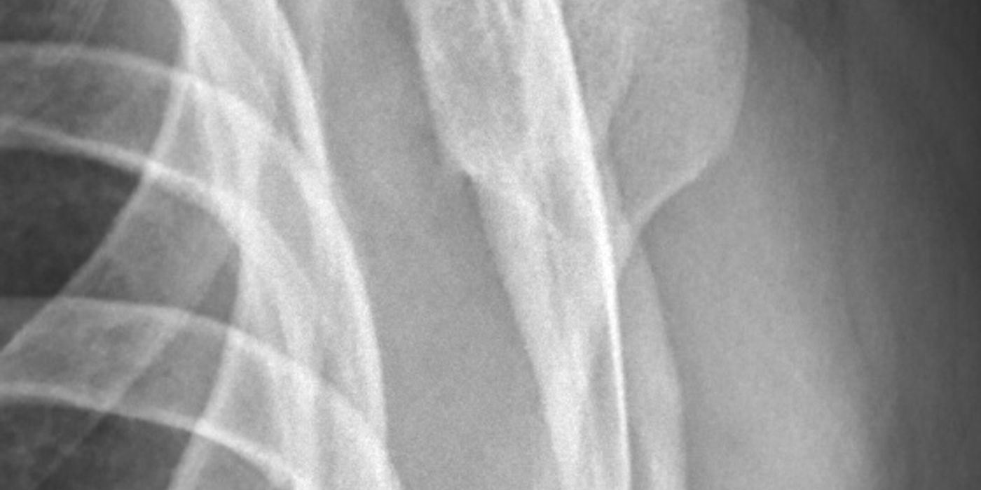
Shoulder dislocation is complete loss of contact between the humeral head and the glenoid, most commonly anteriorly after a fall onto the outstretched, abducted and externally rotated arm. Shoulder instability is the resulting condition — recurrent dislocation, subluxation or apprehension caused by damage to the joint's stabilisers, classically an avulsion of the anteroinferior labrum (Bankart lesion). Management is closed reduction with neurovascular assessment before and after, early mobilisation rather than prolonged immobilisation, and selective stabilisation surgery (arthroscopic Bankart repair, or a Latarjet procedure when there is significant glenoid bone loss) for those at high risk of recurrence.
What anatomy keeps the glenohumeral joint stable?
The glenohumeral joint trades constraint for mobility: the glenoid covers only a small portion of the humeral head, so stability depends on soft tissues. The BESS/BOA care pathway divides these into:
- Static stabilisers — articular conformity, the glenoid labrum (which deepens the socket and increases contact area) and the glenohumeral ligaments. The inferior glenohumeral ligament complex is the principal restraint to anterior translation in abduction–external rotation, which is why it fails in the classic injury mechanism.
- Dynamic stabilisers — concavity-compression from the rotator cuff, the scapular rotators, proprioception and the long head of biceps.
The axillary nerve (C5–6, posterior cord) passes through the quadrilateral space and winds around the surgical neck of the humerus, directly in harm's way during dislocation; regimental badge sensation and deltoid function must be examined and documented. Traumatic anterior dislocation is the most common large-joint dislocation (incidence roughly 8–24 per 100,000 per year), with peaks in young men aged 15–29 and in elderly women.
How does a shoulder dislocation present and how is it diagnosed?
- History: mechanism, age at first dislocation, hand dominance, sport/occupation, number of previous episodes, and symptoms of ongoing instability — these drive later surgical decision-making.
- Examination: loss of the deltoid contour with the arm held in slight abduction; document axillary nerve function (regimental badge sensation, deltoid contraction) and distal neurovascular status before any reduction attempt. The Dec 2021 BOAST on Peripheral Nerve Injury requires nerve function to be assessed and documented "at the first opportunity after injury" and before and after any intervention, in enough detail to compare with later examinations.
- Imaging: the BESS/BOA pathway mandates two radiographic views — an anteroposterior view plus an axial view (or a modified axial/lateral scapular view if pain prevents positioning) — to confirm dislocation and, crucially, its direction, and to identify fractures before reduction.
How is a first-time anterior dislocation managed?
Follow the BESS/BOA Patient Care Pathway (Shoulder & Elbow, 2015), the reference UK standard:
- Reduce in a controlled environment. The principles of safe reduction are consent, adequate analgesia with or without sedation (occasionally general anaesthesia in muscular patients), avoidance of rotational force, post-reduction radiographs in two planes, and re-examination and documentation of neurological and circulatory status.
- Nerve deficit changes the urgency. Where a nerve injury accompanies a dislocation, the Dec 2021 BOAST states "the joint should be reduced immediately". A deficit that appears or worsens after reduction needs prompt specialist referral; as good practice derived from the BESS/BOA pathway, a deficit with no clinical or electromyographic recovery by 6 weeks warrants referral to a peripheral nerve injury service.
- Sling briefly, mobilise early. Immobilisation beyond one week does not reduce recurrence; patients should mobilise as comfort allows. External rotation bracing is not recommended — trial evidence is equivocal and it adds cost and poor compliance for no benefit over a simple sling.
- Rehabilitation: the UK ARTISAN trial (BMJ 2024) randomised 482 adults after first-time traumatic anterior dislocation and found that a programme of additional physiotherapy was not superior to a single session of advice with the option to self-refer, using the Oxford Shoulder Instability Score at 6 months. Routine referral of every first-time dislocator for a full physiotherapy course is therefore not mandatory; structured advice with self-referral is acceptable.
- Screen the cuff in older patients. Around 40% of patients over 40 have a rotator cuff tear after anterior dislocation. The pathway advises routine ultrasound or MRI of the cuff in patients aged 40–60, and an acute traumatic cuff tear is a "red flag" needing urgent specialist referral — early repair gives better results.
What injuries are associated with anterior dislocation?
Figures below are from Robinson et al. (JBJS Am 2012) and the BESS/BOA pathway.
| Injury | Frequency | What to do |
|---|---|---|
| Bankart lesion (± bony Bankart) | Most first dislocations | MR arthrography to define; repair if stabilisation indicated; CT best quantifies glenoid bone loss |
| Hill–Sachs lesion | Almost all dislocations | Usually no treatment; assess engagement/glenoid track if recurrent instability |
| Nerve injury (axillary in two-thirds) | ~13.5% clinically; up to 48% on EMG | Document, reduce promptly, physiotherapy; EMG at 6 weeks if no recovery, then nerve centre referral |
| Greater tuberosity fracture | ~16% | Most reduce with the joint; fix if >5 mm displaced post-reduction; image weekly for 4 weeks for late displacement |
| Rotator cuff tear | ~10% clinically relevant; ~40% over age 40 | Ultrasound/MRI in over-40s; early surgical repair of traumatic tears |
Who needs surgery for recurrent instability — and Latarjet or soft tissue repair?
Recurrence risk is inversely related to age: in the Edinburgh data used by the BESS/BOA pathway, males under 20 have roughly a 72–86% probability of recurrent instability within two years, while Hovelius's 25-year prospective study (JBJS Am 2008) showed many older first-time dislocators never redislocate. Nearly 90% of recurrences occur within two years.
- Primary stabilisation: the pathway supports primary arthroscopic Bankart repair in young, high-demand patients — particularly young men in contact sport or overhead occupations — where trial and Cochrane evidence shows a large relative risk reduction in recurrence versus conservative care.
- Soft tissue (anatomic) repair: arthroscopic repair with suture anchors is as effective as open repair, with recurrence around 8% at 2–4 years. Non-anatomic procedures (e.g. Putti-Platt) cause stiffness and early osteoarthritis and should not be used.
- Latarjet (coracoid transfer): per the BESS/BOA pathway, an open Latarjet should be considered with ≥20% glenoid bone loss or after failed soft tissue stabilisation — not as a routine primary procedure, because recurrence of 2.9–8% is bought at a 25–30% complication rate (non-union, nerve injury, infection).
- Engaging Hill–Sachs defects may be managed with remplissage (infraspinatus capsulotenodesis into the defect) combined with Bankart repair, or with a Latarjet; the modern "glenoid track" concept helps identify off-track lesions needing this. As good practice, the Instability Severity Index Score (Balg & Boileau, JBJS Br 2007) — age ≤20, sport type, hyperlaxity, Hill–Sachs and glenoid bone loss on radiographs — flags patients (score >6) at high risk of failure after isolated arthroscopic repair.
How is posterior dislocation different?
Posterior dislocations are rare (a few per cent of shoulder dislocations) and classically follow seizure, electrocution or high-energy trauma. The arm is locked in internal rotation with blocked external rotation; the AP radiograph may look deceptively normal apart from the "light bulb" sign of the internally rotated head, which is why the pathway's insistence on a second (axial) view matters — an unreduced dislocation of any direction is a red flag requiring same-day emergency referral, and posterior dislocations are notoriously missed at first presentation. As good practice: obtain an axial view or CT, reduce under adequate anaesthesia, and manage the reverse Hill–Sachs (McLaughlin) defect on its size — small defects with a stable reduction can be treated in a sling in neutral/external rotation, larger defects may need subscapularis or lesser tuberosity transfer into the defect (McLaughlin-type procedures), and chronic locked dislocations with major head destruction may need arthroplasty. Bilateral posterior dislocation after a seizure is a classic examination scenario.
What are the complications and prognosis?
- Recurrent instability — the dominant complication in the young; drives most surgery.
- Axillary nerve palsy — most recover; refer to a nerve injury centre if no recovery at 6 weeks.
- Stiffness and post-traumatic osteoarthritis — associated with recurrent instability and with non-anatomic repairs.
- Missed cuff tear — the key cause of persistent weakness in the over-40s ("the young redislocate, the old tear their cuff").
Key points
- Document neurovascular status before and after reduction; two radiographic views before and after, always confirming direction.
- If there is a nerve deficit, reduce the joint immediately (Dec 2021 BOAST on Peripheral Nerve Injury).
- Sling for comfort only — immobilisation beyond one week does not reduce recurrence; external rotation bracing is not recommended.
- ARTISAN (BMJ 2024): structured advice with self-referral is as good as a routine physiotherapy programme after first dislocation.
- Recurrence is age-driven: consider primary arthroscopic Bankart repair in young, high-demand patients; recurrence after modern repair is ~8%.
- Latarjet for ≥20% glenoid bone loss or failed soft tissue surgery; remplissage or Latarjet for engaging Hill–Sachs lesions.
- Image the rotator cuff in patients over 40; repair traumatic tears early.
- Posterior dislocation: seizure/electrocution, locked internal rotation, light-bulb sign — always get the axial view.