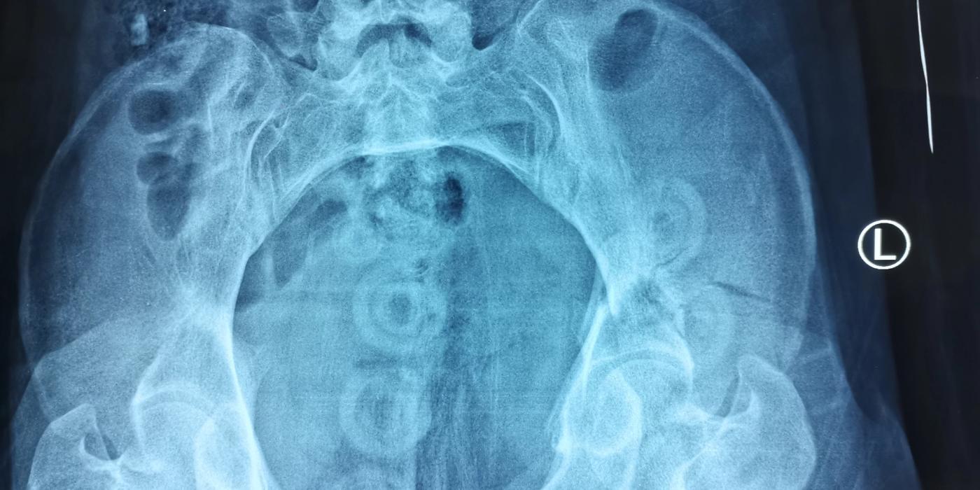
A pelvic ring injury is a disruption of the bony and ligamentous ring formed by the two innominate bones and the sacrum, usually caused by high-energy blunt trauma such as road traffic collisions, falls from height or crush injuries. Because the pelvis is a true ring, displacement at one point almost always implies a second injury elsewhere in the ring, and the same forces that break the ring can tear the adjacent venous plexus, internal iliac branches, bladder and urethra. Management in the UK follows the January 2018 BOAST on the Management of Patients with Pelvic Fractures: early binder application, tranexamic acid, contrast-enhanced CT, haemorrhage control by packing or embolisation, and transfer to a specialist pelvic unit for definitive fixation.
How are pelvic ring injuries classified?
The Young–Burgess classification (Young, Burgess et al., Radiology, 1986) groups injuries by the direction of the causative force. It is the most useful system at the trauma call because it predicts both the resuscitation problem and the reduction manoeuvre: anteroposterior compression injuries open the pelvic volume and bleed, so they respond to circumferential compression; lateral compression injuries are already internally rotated, so aggressive binder tightening adds little and can over-reduce the ring.
| Type | Mechanism and pattern | Stability |
|---|---|---|
| APC I | Anteroposterior force; symphyseal diastasis <2.5 cm, anterior sacroiliac ligaments intact | Stable |
| APC II | Diastasis >2.5 cm; anterior sacroiliac, sacrospinous and sacrotuberous ligaments disrupted ("open book") | Rotationally unstable, vertically stable |
| APC III | Complete sacroiliac disruption with lateral displacement | Rotationally and vertically unstable; highest haemorrhage risk |
| LC I | Lateral force; pubic rami fractures with ipsilateral sacral compression fracture | Usually stable |
| LC II | Rami fractures with iliac wing ("crescent") fracture | Rotationally unstable |
| LC III | Lateral compression on one side with contralateral open-book injury ("windswept pelvis") | Unstable |
| VS | Vertical shear; vertical displacement of the hemipelvis through rami and sacroiliac joint or sacrum | Rotationally and vertically unstable |
| CM | Combined mechanism | Variable |
The Tile classification (A: stable; B: rotationally unstable; C: rotationally and vertically unstable) is the common alternative and maps broadly onto the same concepts.
How do pelvic ring injuries present and how are they diagnosed?
Suspect a pelvic ring injury in any high-energy blunt trauma, in hypotension without an obvious external source, and in the frail patient after a fall who cannot mobilise. Assessment is part of a standard ATLS/CT primary survey:
- Do not "spring" the pelvis. It is unreliable and may dislodge clot; this is established good practice rather than a written standard.
- Examine the perineum, genitalia and rectum and document the findings — an explicit standard of the Aug 2016 urological trauma BOAST for every high-energy trauma patient. Look for blood at the urethral meatus, scrotal or perineal bruising, and wounds of the perineum, buttock or rectum that would make the fracture an open injury.
- Image with contrast-enhanced CT. The Jan 2018 BOAST requires that severely injured patients with high-energy trauma have a "CT scan with IV contrast including head, chest, abdomen and pelvis on admission". NICE NG37 (Fractures (complex): assessment and management) similarly advises against log rolling before pelvic imaging in suspected pelvic fracture unless needed for the airway or a suspected penetrating injury.
- Repeat an X-ray after binder removal. A well-applied binder can perfectly reduce and therefore mask an unstable ring injury, so the BOAST requires a post-binder-removal radiograph in polytrauma patients even when the CT appeared normal.
What is the initial management of a pelvic ring injury?
Initial management is haemorrhage control, anchored to the Jan 2018 BOAST:
- Pelvic binder: apply a properly positioned binder — centred over the greater trochanters, not the iliac crests — whenever active bleeding is suspected, ideally prehospital. NICE NG37 prefers a purpose-made binder, with an improvised binder only if one does not fit.
- Tranexamic acid: give intravenous TXA "as soon as possible and ideally within an hour of injury", and activate the massive transfusion protocol in shocked patients, resuscitating with blood products rather than crystalloid.
- Triage to a Major Trauma Centre: haemodynamically unstable patients with suspected pelvic fractures should go directly to an MTC; if they present to a trauma unit, they are resuscitated and transferred immediately for bleeding control.
- Damage-control surgery: any polytrauma patient going for damage-control laparotomy must have pelvic imaging first, and the binder stays on through surgery until the patient is haemodynamically stable.
- Binder duration: networks must have a binder-removal protocol; the binder should ideally be off within 24 hours of injury (echoed by NICE NG37), with a plan agreed with a pelvic surgeon for a mechanically unstable ring before it comes off.
- Open pelvic fractures: wounds of the lower abdomen, groin, buttock, perineum, anus or rectum need urgent consultant general/colorectal assessment and debridement, with a defunctioning stoma considered for anorectal and some perineal wounds.
What if the patient remains unstable despite the binder?
For active pelvic bleeding unresponsive to resuscitation, the BOAST requires either extraperitoneal pelvic packing or interventional radiology with selective arterial embolisation, and mandates that every MTC has an established protocol for choosing between them. In practice, packing suits the patient in extremis or already in theatre (most pelvic bleeding is venous or bony), whereas angioembolisation suits arterial blush on CT in a patient stable enough to reach the IR suite. REBOA (resuscitative endovascular balloon occlusion of the aorta, zone III) has been used as a bridge in exsanguinating pelvic haemorrhage, but the UK-REBOA randomised trial (Jansen et al., JAMA, 2023) found higher 90-day all-cause mortality with an ED REBOA strategy than with standard care alone (54% vs 42%) and was stopped early; it should be described at interview as unproven and potentially harmful outside selected protocols, not as standard UK care.
How are urological injuries managed?
Bladder and urethral injury must be actively suspected, diagnosed and managed according to the Aug 2016 BOAST on Urological Trauma Associated with Pelvic Fractures:
- "A single, gentle attempt at catheterization, by an experienced doctor, is permissible" — a 16F soft silicone catheter in adults, with the findings documented.
- If the catheter will not pass or drains only blood, "do NOT inflate balloon" — withdraw it and perform a retrograde urethrogram. Blood-stained urine in a passed catheter warrants retrograde cystography.
- Failed urethral catheterisation mandates a percutaneous suprapubic catheter by Seldinger technique under ultrasound guidance, 3–4 finger-breadths above the symphysis — sited high to preserve the surgical field, and discussed early with the pelvic fracture service because it can affect fixation timing.
- Intraperitoneal bladder rupture needs emergency laparotomy and repair; extraperitoneal rupture is usually managed with catheter drainage, but primary repair is recommended when an unstable fracture is being fixed, and bladder neck rupture always requires primary repair.
- For complete urethral rupture in adult males the recommended definitive treatment is delayed repair at three months in a reconstructive centre; acute primary re-alignment during fracture surgery is not recommended, as reduction of the pelvis indirectly re-aligns the urethra.
What is the definitive management?
Temporary mechanical stabilisation — anterior external fixation, and skeletal traction for displaced vertical shear patterns — is used when early definitive surgery is not possible. The Jan 2018 BOAST then sets two timelines: trauma unit patients needing surgical stabilisation must be "referred and safely transferred to a specialist centre within 24 hours", and pelvic ring reconstruction should take place "within 72 hours of the stabilisation of the patient's physiological state if associated injuries allow". Typical constructs are symphyseal plating for anterior disruption and percutaneous iliosacral or trans-sacral screws for the posterior ring, with open posterior fixation for irreducible or highly unstable patterns; the choice is a specialist-centre decision. Displaced low-energy fragility fractures of the pelvis that prevent mobilisation should also be discussed with the specialist centre, since fixation can restore independence in older patients. A written thromboprophylaxis policy, TARN data submission and specialist follow-up complete the pathway.
What are the complications and prognosis?
- Early: exsanguinating haemorrhage, open fracture contamination and pelvic sepsis, urological injury, lumbosacral plexus and L5/S1 nerve root injury, and a high rate of venous thromboembolism.
- Late: malunion and leg-length discrepancy, chronic pelvic and sacroiliac pain, urethral stricture, and sexual dysfunction — common after displaced anterior ring injuries. The BOASTs require written information on sexual dysfunction for all potentially sexually active patients and access to andrological services at MTCs.
- Prognosis tracks the injury pattern and physiology at presentation: stable LC I/APC I injuries usually do well with protected weight-bearing, whereas unstable posterior ring injuries carry significant mortality from haemorrhage and long-term functional, urological and psychological sequelae, which is why BOAST mandates follow-up in a specialist pelvic or rehabilitation clinic.
Key points
- Treat the pelvis as a ring: one break usually means two, and the injury pattern (Young–Burgess) predicts bleeding risk and management.
- Binder on early and correctly positioned, TXA within an hour, blood-product resuscitation, and direct triage of unstable patients to an MTC (Jan 2018 BOAST).
- Ongoing instability despite the binder means packing or angioembolisation by local protocol; UK-REBOA (JAMA 2023) showed no benefit and probable harm from routine ED REBOA.
- Examine and document perineum, genitalia and rectum; one gentle catheter attempt only, never inflate the balloon against resistance, retrograde urethrogram then suprapubic catheter if it fails (Aug 2016 urological BOAST).
- Binder off within 24 hours with a post-removal X-ray; transfer for stabilisation within 24 hours; definitive reconstruction within 72 hours of physiological stabilisation.
- Warn about and follow up sexual dysfunction, urological sequelae and VTE — these are audited standards, not optional extras.