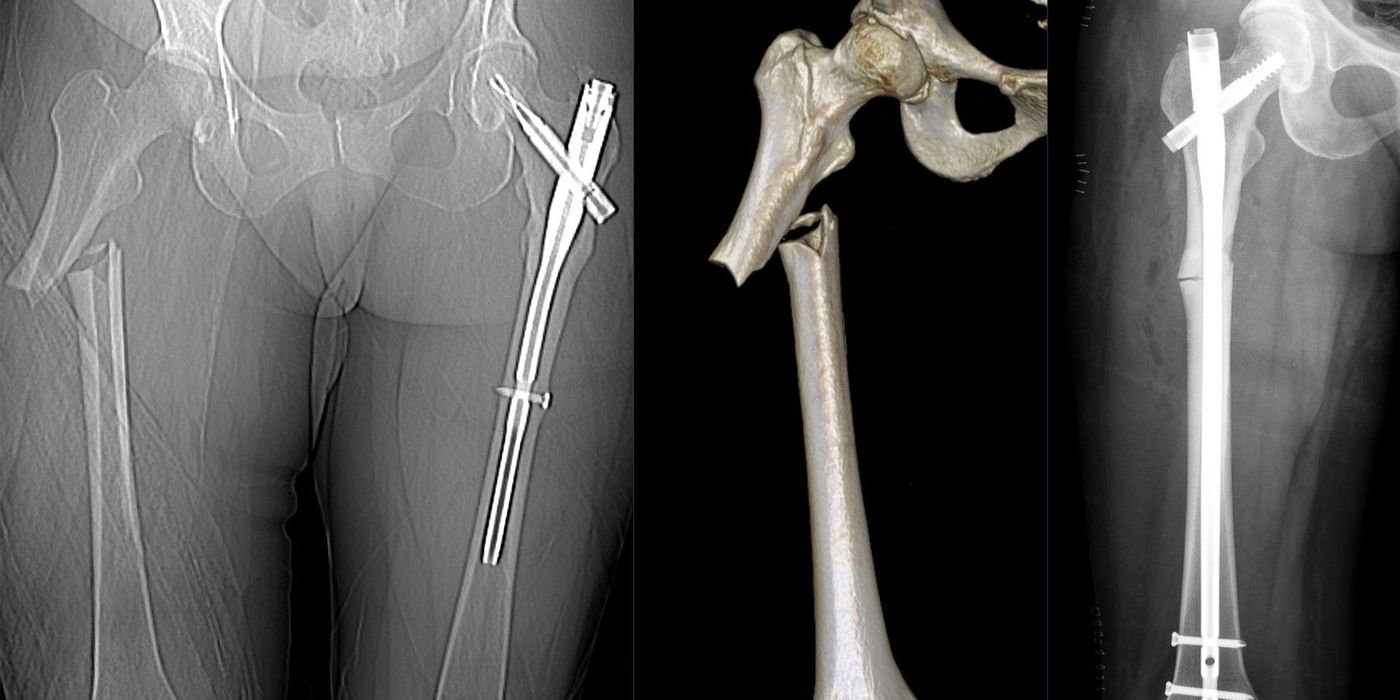
A femoral shaft fracture is a break in the diaphysis of the femur, between the lesser trochanter proximally and the metaphyseal flare of the distal femur. It follows a bimodal pattern: high-energy trauma (road traffic collisions, falls from height) in younger adults, and low-energy fragility, pathological or periprosthetic fractures in older patients. Definitive treatment for most adults is reamed, statically locked intramedullary (IM) nailing, with the timing of surgery dictated by the patient's physiology — early total care in the stable patient, damage control orthopaedics in the unstable or borderline patient.
Who gets femoral shaft fractures, and how are they classified?
In young adults the femur is one of the strongest bones in the body, so a shaft fracture implies major energy transfer and should prompt a search for associated injuries (pelvis, ipsilateral femoral neck, knee ligaments, chest, head). In older patients a fracture after a simple fall is a fragility injury; a fracture with no or minimal trauma should raise suspicion of a pathological cause (metastatic disease — image the whole bone before nailing) or an atypical femoral fracture in long-term bisphosphonate users, classically a transverse lateral-cortex fracture with cortical beaking and prodromal thigh pain.
Fractures are described by the AO/OTA system (femur = bone 3, diaphysis = segment 2: simple 32A, wedge 32B, multifragmentary 32C) and by the Winquist–Hansen grading of comminution (0–IV), which historically guided locking but is now mainly of descriptive value since static locking is routine. Always record open versus closed status and neurovascular findings.
How do they present, and what must the initial assessment include?
The patient presents with a painful, swollen, shortened and externally rotated thigh after trauma. Assessment follows ATLS principles — a femoral shaft fracture is a circulation problem as well as a limb problem: a closed shaft fracture is classically associated with 1,000–1,500 ml of blood loss into the thigh, and bilateral fractures can produce class III–IV shock on their own.
- Catastrophic haemorrhage and shock: manage along major-haemorrhage protocols; give tranexamic acid as soon as possible and within 3 hours of injury where there is active or suspected active bleeding, as per NICE NG39 (Major trauma, 2016).
- Vascular status: as per the Jun 2026 BOAST on arterial injuries, "clinical arterial examination is mandatory and specific findings and timings must be documented". A deformed, pulseless limb requires urgent realignment and splinting with re-examination afterwards. If arterial injury is suspected, "CT angiogram is recommended … and should be performed concurrently with whole-body CT".
- Open wounds: as per the Dec 2017 BOA/BAPRAS BOAST on open fractures, give intravenous prophylactic antibiotics "as soon as possible, ideally within 1 hour of injury", photograph the wound, remove gross contamination only, and cover with a saline-soaked dressing.
- Associated injuries: examine the pelvis, hip and knee. An ipsilateral femoral neck fracture is a well-recognised and easily missed association — dedicated hip imaging (and scrutiny of the trauma CT) before nailing is good practice, and intra-operative screening of the neck after nailing is widely recommended.
- Splintage: NICE NG38 (Fractures: non-complex, 2016) recommends a traction splint — or the adjacent leg as a splint — in the pre-hospital setting for suspected fractures above the knee. Traction reduces pain, bleeding into the thigh and further soft-tissue injury.
What is the emergency management?
- Resuscitate along ATLS lines; treat the fracture as part of "C".
- Analgesia: intravenous opioids plus a femoral nerve or fascia iliaca block (good practice; avoid regional blocks where compartment syndrome of the thigh is a concern, and monitor for it as per the Jul 2025 BOAST on compartment syndrome — rare in the thigh but limb-threatening).
- Apply skin or skeletal traction / a traction splint until definitive surgery.
- If the limb is ischaemic, the Jun 2026 BOAST is explicit: "revascularisation is an emergency procedure (NCEPOD 1) and should be commenced within one hour of arrival to hospital", with haemorrhage controlled "immediately by direct pressure, wound packing, or tourniquet applied as distally as possible". A temporary vascular shunt is recommended to restore flow rapidly, followed by skeletal stabilisation and then definitive vascular repair. Any decision toward early amputation must be made and documented by two consultants.
- Open femoral shaft fractures follow the open-fracture BOAST timelines: immediate debridement for highly contaminated wounds or vascular compromise, "within 12 hours of injury for other solitary high energy open fractures", and within 24 hours for low-energy wounds, delivered by a combined orthoplastic team.
Early total care or damage control orthopaedics — how is timing decided?
This is the core interview discussion. Bone et al. (JBJS Am, 1989), in a prospective randomised study of 178 patients, showed that delayed femoral stabilisation in multiply injured patients increased pulmonary complications (ARDS, fat embolism, pneumonia), ICU days and hospital stay — the foundation of early total care (ETC): definitive reamed nailing within 24 hours in the physiologically stable patient. Subsequent work by Pape and the EPOFF study group (J Trauma, 2003) showed that primary reamed nailing amplifies the systemic inflammatory response (e.g. IL-6) compared with initial external fixation, underpinning damage control orthopaedics (DCO) — temporary spanning external fixation for the unstable or "borderline" patient, converting to a nail once physiology is restored (conversion within days, and generally before two weeks, limits pin-site–related infection risk; good practice).
| Patient category | Typical features | Strategy |
|---|---|---|
| Stable | Normal physiology, responding to resuscitation, no lethal-triad features | Early total care: reamed IM nail within 24 h |
| Borderline | e.g. ISS > 40, significant chest injury, bilateral femoral fractures, hypothermia, rising lactate, moderate head injury | Reassess after resuscitation; low threshold for DCO |
| Unstable / in extremis | Persistent shock, coagulopathy, acidosis, hypothermia (the "lethal triad") | Damage control: rapid external fixation, haemorrhage control, ICU; delayed nailing |
The decision is physiological, not radiographic, and is made jointly with the anaesthetic/ICU team using trends in lactate, base excess, temperature and coagulation rather than a single snapshot.
What is the definitive management?
The gold standard for adult femoral shaft fractures is an antegrade, reamed, statically locked intramedullary nail, which allows early mobilisation and, in most patterns, immediate full weight bearing. The evidence for reaming is robust: the Canadian Orthopaedic Trauma Society multicentre RCT (JBJS Am, 2003; 224 patients) reported nonunion in 1.7% after reamed nailing versus 7.5% unreamed. Retrograde nailing is a recognised alternative for ipsilateral neck-and-shaft injuries (the neck takes fixation priority), floating knee, bilateral fractures, obesity or pregnancy. Plate fixation is reserved for narrow or obliterated canals, peri-implant and periprosthetic fractures, and some adolescent injuries; definitive external fixation is rarely used in adults in the UK.
For open fractures, the BOAST sequencing applies verbatim: "definitive internal stabilisation should only be carried out when it can be immediately followed with definitive soft tissue cover", and definitive cover should be achieved within 72 hours of injury if not done at first debridement.
Bilateral femoral shaft fractures
Bilateral fractures mark massive energy transfer and carry higher rates of ARDS and mortality than unilateral injuries. Treat the patient as at least borderline: many will be best served by damage control with staged nailing, while single-sitting bilateral nailing is reasonable only in the genuinely stable patient with an experienced team and explicit anaesthetic agreement (good practice).
Periprosthetic femoral shaft fractures
Fractures around a hip stem or total knee replacement are classified by the Vancouver system / Unified Classification System: with a well-fixed stem (B1) fixation is appropriate; a loose stem (B2/B3) generally needs revision arthroplasty, often combined with fixation. These are predominantly frailty injuries: as per the BOAST on the older or frail orthopaedic trauma patient, surgery "should be performed to allow full weight-bearing for activities required for daily living and within 36 hours of admission, in line with current hip fracture care", with a Comprehensive Geriatric Assessment commencing within 72 hours and complex periprosthetic patterns discussed with a specialist unit.
What are the complications and prognosis?
- Fat embolism syndrome: typically 24–72 hours after injury, with the triad of hypoxaemia, neurological deterioration and a petechial rash. Diagnosis is clinical, using Gurd and Wilson's criteria (JBJS Br, 1974) — broadly two major criteria, or one major plus four minor. Treatment is supportive (oxygen, ventilation, resuscitation); early fracture stabilisation reduces incidence, as shown by Bone et al.
- Nonunion and malunion: nonunion is uncommon after reamed nailing (under 2% in the COTS trial); malrotation is the commonest technical error — compare rotation with the contralateral limb intra-operatively (good practice).
- Infection: uncommon in closed injuries; suspected fracture-related infection should be investigated and managed along the principles of the Sep 2019 BOAST on fracture-related infections.
- Missed ipsilateral femoral neck fracture, thigh compartment syndrome, and neurovascular injury — all rare but repeatedly examined, and all prevented or mitigated by the structured assessment above.
Prognosis after isolated fracture is good: most nailed fractures unite within three to six months with return to pre-injury function, while outcomes in polytrauma are driven by the associated injuries.
Key points
- A femoral shaft fracture is a marker of major energy transfer in the young and a fragility, pathological or periprosthetic injury in the old.
- Assess along ATLS lines: expect 1,000–1,500 ml blood loss, document pulses (arterial BOAST, Jun 2026), splint in traction (NICE NG38), give antibiotics within 1 hour if open (BOAST, Dec 2017).
- Definitive treatment is a reamed, statically locked IM nail — reaming reduces nonunion (COTS RCT, 2003).
- Timing is physiological: early total care in the stable patient (Bone, 1989); damage control external fixation in the borderline or unstable patient (Pape/EPOFF, 2003).
- Bilateral fractures are a borderline-at-minimum situation; periprosthetic fractures in the frail follow the 36-hour, full-weight-bearing frailty BOAST standard.
- Know fat embolism syndrome cold: 24–72 hours, hypoxia–confusion–petechiae, Gurd and Wilson criteria, supportive care, prevented by early stabilisation.