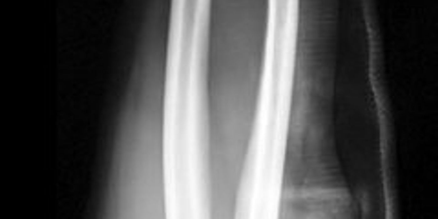
Forearm fractures are breaks of the radius and/or ulna between the elbow and the wrist, including both-bone (diaphyseal) fractures and the fracture-dislocation patterns of Monteggia (ulna fracture with radial head dislocation) and Galeazzi (radius fracture with distal radio-ulnar joint disruption). In adults the forearm behaves as a joint — the two bones rotate around each other in pronosupination — so displaced diaphyseal fractures are treated like intra-articular injuries, with anatomical open reduction and plate fixation. When the fracture is open, management follows the national open fracture pathway: intravenous antibiotics within an hour, urgent combined orthopaedic and plastic surgical (orthoplastic) care, timely debridement, and definitive fixation with soft tissue cover within 72 hours.
What injury patterns matter in the forearm?
- Both-bone (diaphyseal) fractures — usually high-energy in adults (road traffic collisions, falls from height, direct blows). Loss of the radial bow or of interosseous space costs forearm rotation.
- Monteggia fracture-dislocation — proximal-third ulna fracture with dislocation of the radial head, classified by Bado (Clin Orthop, 1967). The radial head almost always reduces once ulnar length and alignment are anatomically restored; a radial head that will not reduce suggests malreduction of the ulna or interposed tissue (commonly annular ligament).
- Galeazzi fracture-dislocation — radial shaft fracture (classically at the junction of the middle and distal thirds) with disruption of the distal radio-ulnar joint (DRUJ). Known as the "fracture of necessity" because non-operative treatment in adults reliably fails.
- Isolated ulna ("nightstick") fracture — a direct-blow injury; minimally displaced fractures can be treated non-operatively, but a Monteggia pattern must be actively excluded on elbow radiographs first.
- Open fractures — the subcutaneous ulnar border means even low-energy injuries can be open. Grade with the Gustilo–Anderson classification (JBJS Am 1976; IIIA/B/C subdivision, J Trauma 1984) after debridement, not in the emergency department.
| Bado type | Radial head dislocation | Ulna fracture |
|---|---|---|
| I | Anterior | Diaphysis, anterior angulation (most common) |
| II | Posterior | Diaphysis, posterior angulation |
| III | Lateral | Metaphysis (typically children) |
| IV | Anterior | Both radius and ulna fractured |
How should a forearm fracture be assessed?
Assessment follows ATLS principles in high-energy injury; the limb examination must be systematic and documented.
- Neurovascular examination — document radial and ulnar pulses, capillary refill, and median (including anterior interosseous), ulnar and radial/posterior interosseous nerve function. The peripheral nerve injury BOAST (Dec 2021) requires examination "at the first opportunity after injury" and repetition "after any intervention to the limb such as injection, manipulation or application of cast", recorded in enough detail for confident comparison later.
- Vascular concern — as per the Jun 2026 BOAST on arterial injuries, clinical arterial examination is mandatory with findings and timings documented; if arterial injury is suspected, CT angiography is recommended and revascularisation is an NCEPOD 1 emergency that "should be commenced within one hour of arrival to hospital".
- Radiographs — orthogonal views of the whole forearm including the elbow and wrist joints. On every view, check that the radiocapitellar line passes through the capitellum (misses = Monteggia) and inspect the DRUJ for widening, ulnar styloid fracture or ≥5 mm radial shortening (Galeazzi).
- The wound and the compartments — inspect once, photograph, remove gross contamination only, then seal the wound; and actively assess for compartment syndrome (below), to which the forearm is notably vulnerable.
How is an open forearm fracture managed initially?
Apply the BOA/BAPRAS Open Fractures standard (BOAST 4, Dec 2017) and NICE NG37 (Fractures — complex), which are aligned:
- Antibiotics — intravenous prophylactic antibiotics as soon as possible and ideally within 1 hour of injury; if not given pre-hospital, give immediately on arrival. Check and record tetanus status (good practice).
- Assess, photograph, dress — document neurovascular status before and after any reduction or splintage; handle the wound only to remove gross contaminants, photograph it, then dress with saline-soaked gauze under an occlusive film. Do not irrigate open long-bone fractures in the pre-hospital or emergency department setting, and do not repeatedly uncover the wound.
- Realign and splint — restore gross alignment in an above-elbow backslab and re-examine.
- Orthoplastic referral — open long-bone fractures should be managed by a combined orthopaedic–plastic surgery service; discuss and transfer according to the network pathway rather than performing an incomplete "washout" locally out of hours.
- Debridement timing (per BOAST 4 / NG37): immediately for highly contaminated wounds (agricultural, aquatic, sewage) or when there is vascular compromise; within 12 hours for other solitary high-energy open fractures; within 24 hours for low-energy open fractures. Debridement should use extensile incisions compatible with fasciotomy approaches, performed by senior orthopaedic and plastic surgeons together.
What is the definitive management?
Closed injuries
- Displaced both-bone fractures in adults — anatomical open reduction and compression plate fixation of both bones (typically 3.5 mm dynamic compression plates through separate approaches: volar Henry for the radius, direct subcutaneous approach for the ulna). Compression plating gives union rates in excess of 96% (Anderson et al., JBJS Am 1975). Restore radial bow; check pronosupination and DRUJ stability on table.
- Monteggia — anatomical plate fixation of the ulna, then confirm concentric radial head reduction on screening. If it does not reduce, revisit the ulnar reduction before opening the radiocapitellar joint.
- Galeazzi — anatomical plate fixation of the radius, then reassess the DRUJ: if stable, early mobilisation; if reducible but unstable, immobilise in supination with or without DRUJ pinning; if irreducible, open the DRUJ (interposed extensor carpi ulnaris is the classic block) — labelled good practice rather than guideline-mandated.
- Isolated minimally displaced ulnar shaft fractures — functional brace or cast with early movement; operate for displacement >50%, angulation >10°, or any joint involvement (good practice thresholds).
Open injuries
- At first debridement, if the wound allows primary closure, definitive internal fixation and closure at the same sitting is standard. Otherwise stabilise (spanning external fixation or, in the forearm, often definitive plating with temporary coverage) and achieve definitive fixation and soft tissue cover within 72 hours of injury, as per BOAST 4/NG37. Use fresh instruments and re-prep between debridement and fixation.
- Irrigation at debridement: the FLOW trial (NEJM, 2015) found very-low-pressure saline irrigation acceptable and soap solution associated with more reoperations — normal saline without additives is the evidence-based choice.
- Nerve injury with a penetrating or lacerating wound requires discussion with a specialist nerve injury service within 24 hours (Dec 2021 BOAST); explore the nerve at the time of debridement where expertise allows.
- If revascularisation is needed, use a temporary vascular shunt, stabilise the skeleton after reperfusion, and remember the Jun 2026 BOAST instruction that "fasciotomies must always be considered after revascularisation".
How is forearm compartment syndrome recognised and managed?
The forearm is a classic site, particularly after crush injuries, high-energy both-bone fractures and revascularisation. As per the Jul 2025 BOAST on compartment syndrome:
- Suspect it with pain disproportionate to the injury and pain on passive stretch of the digits; high-risk patients need hourly documented assessments, and escalating analgesic requirement is a red flag — the dose and timing of opiates must be recorded.
- Immediately split and release circumferential dressings and casts, elevate to heart level, and reassess within 30 minutes.
- In patients who cannot be assessed clinically (unconscious, blocks), use compartment pressure monitoring: a differential pressure (diastolic minus compartment) below 30 mmHg indicates increased risk — the threshold derived from McQueen and Court-Brown (JBJS Br, 1996).
- "Immediate surgical decompression should follow a diagnosis of compartment syndrome." In the forearm this means an extensile volar fasciotomy releasing the superficial and deep volar compartments and the carpal tunnel, adding a dorsal release where needed (good practice technique). Plan soft tissue management with plastic surgery and return for re-look within 48–72 hours; in late presentations (complete, established syndrome), a documented two-consultant decision may favour non-operative management with renal protection.
What are the complications and prognosis?
- Malunion and loss of rotation — failure to restore the radial bow limits pronosupination.
- Non-union — uncommon after anatomical compression plating; manage established aseptic non-union with compression plating and bone graft.
- Radio-ulnar synostosis — risk rises with high-energy injury, head injury and a single approach to both bones; excise once mature if function is limited.
- Infection and fracture-related infection — follow the Sep 2019 BOAST on fracture-related infections: multidisciplinary management, deep sampling before targeted antibiotics.
- Nerve injury — posterior interosseous nerve palsy in Monteggia injuries is usually a neurapraxia that recovers; document serially and escalate per the nerve injury BOAST if it does not.
- Refracture after plate removal — a recognised risk; remove forearm plates only for a clear indication and not before 18 months (good practice).
- Missed compartment syndrome — leads to Volkmann's ischaemic contracture, the most devastating preventable outcome.
Key points
- The adult forearm functions as a joint: displaced diaphyseal fractures need anatomical open reduction and compression plate fixation of both bones.
- Always image and examine the elbow and wrist — an "isolated" shaft fracture may be a Monteggia or Galeazzi injury.
- Open fractures: IV antibiotics within 1 hour; debride immediately if contaminated or dysvascular, within 12 h if high energy, within 24 h if low energy; fixation and definitive cover within 72 h (BOAST 4 / NICE NG37).
- Reduce the joint by fixing the bone: anatomical ulnar reduction reduces the radial head (Monteggia); anatomical radial reduction is the first step to a stable DRUJ (Galeazzi).
- Forearm compartment syndrome demands hourly assessment in high-risk patients, ΔP <30 mmHg as the monitoring threshold, and immediate fasciotomy on diagnosis (Jul 2025 BOAST).
- Suspected arterial injury is an NCEPOD 1 emergency: revascularise within one hour of arrival, shunt then stabilise, and always consider fasciotomy afterwards (Jun 2026 BOAST).