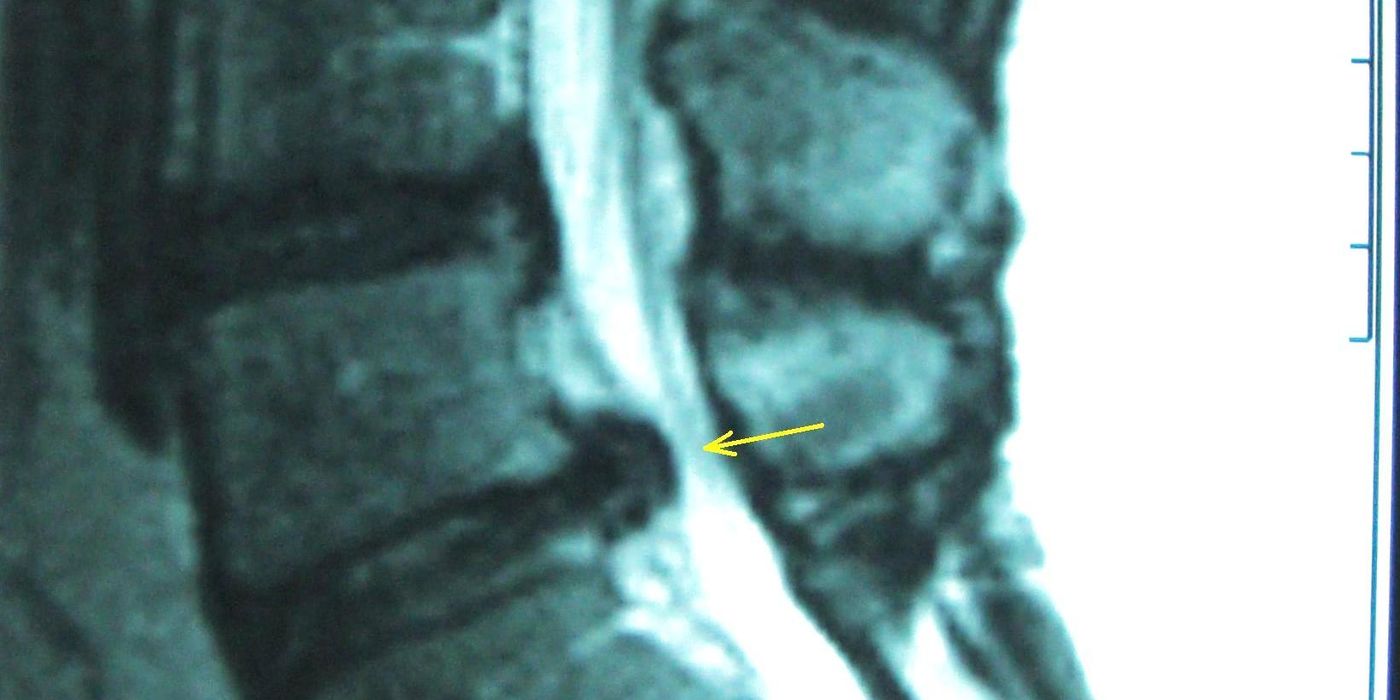
Cauda equina syndrome (CES) is a spinal surgical emergency caused by acute or rapidly progressive compression of the lumbosacral nerve roots in the lumbar or sacral spinal canal, most commonly by a large lumbar disc prolapse. Untreated, it can cause permanent loss of bladder, bowel and sexual function and lower limb paralysis. In the UK it is managed according to the GIRFT National Suspected Cauda Equina Syndrome Pathway (February 2023), which mandates emergency MRI at the presenting hospital and decompression as an NCEPOD emergency.
What is cauda equina syndrome and why does it matter?
The cauda equina is the bundle of lumbosacral nerve roots (L2–S5) below the conus medullaris, carrying motor and sensory supply to the legs, bladder, bowel and perineum. CES is rare — around 1 to 3 per 100,000 population per year — but its consequences are life-changing, and the reliability of clinical diagnosis is low. The commonest cause is a large central lumbar disc prolapse; rarer causes include haematoma, trauma, infection, tumour and complications of spinal or epidural anaesthesia. The February 2023 national pathway, led by GIRFT and co-badged by eleven organisations including the British Association of Spine Surgeons (BASS), the Society of British Neurological Surgeons (SBNS), the BOA and the Royal College of Radiologists, followed a 2021 HSIB national investigation into delayed diagnosis and is now the reference standard for UK practice — and for the ST3 interview.
What are the red flag symptoms of cauda equina syndrome?
Under the GIRFT pathway, a patient with leg pain and/or back pain and recent onset (within two weeks) of any of the following warrants an emergency referral to the nearest hospital with emergency MRI provision:
- difficulty initiating micturition or impaired sensation of urinary flow;
- altered perianal, perineal or genital sensation (S2–S5 dermatomes) — "the area may be small or as big as a horse's saddle", subjectively reported or objectively tested;
- severe or progressive bilateral neurological deficit of the legs, such as major motor weakness of knee extension, ankle eversion or foot dorsiflexion;
- loss of sensation of rectal fullness;
- sexual dysfunction — inability to achieve erection or ejaculate, or loss of vaginal sensation.
Two points are repeatedly examined. First, no single symptom or sign is pathognomonic, no combination has good diagnostic accuracy, and negative physical tests do not rule out CES if positive subjective symptoms are present. Second, the pathway created a distinct urgent (two-week) route: sudden-onset bilateral radicular leg pain, or unilateral radicular pain progressing to bilateral, without CES symptoms, is a warning presentation referred urgently to an MSK triage service with explicit safety-netting (warning card and video); any deterioration converts it to an emergency referral. A digital rectal examination is not necessary for referral, but subjective perianal sensation must be recorded, and every assessment must be documented with times, dates and progression — this documentation is what stands up medicolegally.
What is the difference between CESS, CESI and CESR?
Severity at presentation is the strongest determinant of prognosis and drives surgical priority:
| Stage | Definition | Implication |
|---|---|---|
| CESS (suspected) | Red flag symptoms without established sphincter dysfunction | Emergency MRI to confirm or exclude compression |
| CESI (incomplete) | Neurogenic urinary symptoms (altered sensation, difficulty initiating or controlling flow) with retained executive bladder control | Best prognosis if decompressed before progression — operate "as quickly as possible" |
| CESR (retention) | Painless urinary retention with overflow incontinence | Poorer prognosis, but around 70% still benefit from decompression |
The window that matters is the transition from CESI to CESR: this is the deterioration that surgery aims to prevent, which is why CESI is treated with the greatest urgency.
How is suspected cauda equina syndrome assessed in hospital?
On arrival in secondary care the patient is triaged, examined and a bladder scan performed — but the pathway is emphatic about its role: "Bladder scans should NOT be used in isolation or as a discriminator in deciding to request an MRI or undertake emergency surgery." In the UCES prospective cohort (Woodfield et al., 2023), 60% of patients who underwent emergency decompression for CES had a post-void residual (PVR) below 200 ml — a normal scan does not exclude CES. Used correctly, the scan adds information:
- if the patient is able to void, document pre-void volume and PVR; a PVR >200 ml in suspected CES makes the diagnosis around 20 times more likely;
- if the patient is unable to void, scan and, if the volume exceeds 600 ml, catheterise (documenting whether the catheter tug is sensate) to prevent bladder distension injury.
Keep the patient nil by mouth from the point of requesting emergency imaging, in case emergency surgery follows.
When is emergency MRI needed and how quickly?
MRI is the critical diagnostic investigation, and the 2023 pathway sets hard operational standards: MRI at the presenting hospital is best practice, in and out of hours, and an emergency MRI for suspected CES "should be undertaken as soon as possible, and certainly within four hours of request to radiology", with local provision required by June 2024. Key rules, several of which reverse older habits:
- discussion with the on-call spinal surgical service is not required before the MRI and "may lead to unwarranted delay" — echoing the SBNS/BASS 2018 standards, which state the MRI "must be undertaken as an emergency in the patient's local hospital and a diagnosis achieved prior to any discussion with the spinal services";
- the request should be discussed with a senior decision-maker (ST4 or equivalent, or above/consultant) before referral to radiology;
- the emergency scan takes precedence over all routine and elective cases;
- a screening sagittal T2 sequence is sufficient to demonstrate or exclude cauda equina compression, with further sequences only if compression is found; if no compression is seen, a sagittal T2 of the cervical and thoracic spine is added;
- if MRI is absolutely contraindicated, CT or CT myelography may provide satisfactory imaging;
- on-call surgical teams may review out-of-hours images before the formal radiology report, which should be available within one hour.
Three imaging outcomes follow: compression confirmed (immediate referral to the spinal surgical service, patient nil by mouth, Category 2 ambulance transfer if needed); no cauda equina compression but neural compression explaining sciatica (safety-net and refer to MSK triage); or no cause found (consider alternative diagnoses, including non-compressive pathology, and referral to other specialties).
How quickly should the cauda equina be decompressed?
Confirmed compression means immediate referral, consent, catheterisation before the start of surgery (avoiding bladder distension injury), registration on the British Spine Registry and a time-stamped, documented review by the surgical team. On timing, the pathway states that surgery for CESI "should be undertaken as quickly as possible, as an NCEPOD E1/E2 emergency. Cauda Equina sits between E1 and E2, as it is time-sensitive and life-changing, but not life-threatening. Any reason for delay should be documented." For CESR, timing is at the operating surgeon's discretion but "should still in this instance be undertaken within 24 hours of MRI imaging". The SBNS/BASS 2018 standards underpin this: "Nothing is to be gained by delaying surgery and [it] should be undertaken at the earliest opportunity, considering the duration and clinical course of symptoms and signs, and the potential for increased morbidity while operating in the night." Laminectomy, hemilaminectomy and laminotomy are all acceptable techniques; trainees must be appropriately supervised; and surgery in which anaesthesia would start between midnight and 07:30 requires consultant on-call sanction.
What are the outcomes, complications and follow-up?
Complication rates in CES decompression are around six times higher than in non-CES decompressive surgery. Post-operatively the pathway mandates early mobilisation (usually day one), a trial without catheter with pre- and post-void bladder scans, and a strict rule: patients who fail TWOC are not re-trialled on the ward — they are re-catheterised, taught intermittent self-catheterisation where possible, and followed up by the regional spinal cord injury or urology service. Bowel care (stimulant plus softener, digital stimulation education per MASCIP guidance), sexual function review (a phosphodiesterase-5 inhibitor is worth trialling in men and women) and psychological support are all mandated, with a six-to-eight-week face-to-face review and referral of any patient with ongoing symptoms to the regional spinal cord injury unit. Even with prompt surgery some patients have permanent deficits, but treating before symptoms become severe reduces the risk of lasting disability.
Why is cauda equina syndrome so important medicolegally?
CES accounted for 23% of litigated claims relating to spinal surgery in England in GIRFT's review of claims between 2013/14 and 2015/16 — the single largest category. Claims typically turn on delayed recognition, delayed imaging and poor documentation rather than surgical technique. This is why the pathway is so prescriptive about recording symptoms with onset, duration, frequency and progression, the time and date of each assessment, who was referred to and when, and the reason for any delay to scanning or surgery. As good practice for the interview: a candidate who safety-nets the "warning sign" patient, scans without waiting for spinal team approval, and documents contemporaneously is demonstrating exactly what the national pathway was written to achieve.
Key points
- CES is acute compression of the lumbosacral nerve roots, usually by a large disc prolapse; it is rare (1–3 per 100,000) but a true surgical emergency.
- Emergency referral is triggered by leg/back pain plus any one red flag of less than two weeks' onset; bilateral sciatica without CES symptoms takes the urgent two-week MSK route with safety-netting.
- Bladder scanning is an adjunct only — 60% of operated CES patients had a PVR under 200 ml; never use it to gatekeep MRI or surgery.
- Emergency MRI at the presenting hospital, as soon as possible and certainly within four hours of request; no prior discussion with the spinal service is required.
- Decompress CESI as an NCEPOD E1/E2 emergency; CESR within 24 hours of MRI; anaesthesia starting between midnight and 07:30 needs consultant sanction.
- Catheterise before surgery, register on the British Spine Registry, and never repeat a failed TWOC — refer to the spinal cord injury or urology service.
- CES is the leading source of spinal litigation (23% of litigated spinal surgery claims); meticulous, time-stamped documentation is part of the treatment.