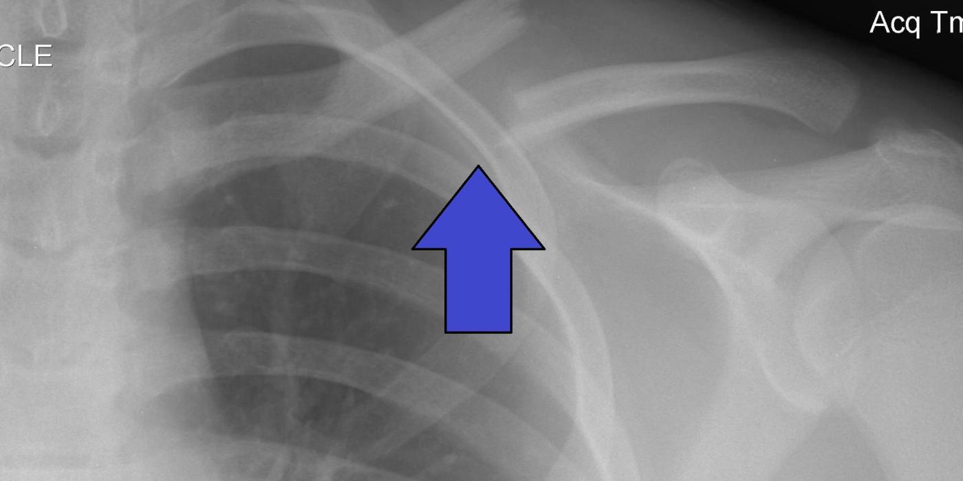
A clavicle fracture is a break in the collarbone, the strut connecting the upper limb to the axial skeleton. It is one of the commonest adult fractures, accounting for around 2.6–4% of all fractures, and roughly 80% occur in the middle third (midshaft). Most heal well with a sling alone, but completely displaced midshaft fractures in adults carry a nonunion risk of around 10–15% if treated non-operatively, so the decision between sling and plate fixation is individualised rather than automatic.
How do clavicle fractures present and how are they diagnosed?
The typical mechanism is a fall onto the point of the shoulder, or a direct blow, during sport, cycling or a road traffic collision; in older patients a simple fall predominates and lateral-end fractures become relatively more common. Patients present with pain, swelling and often visible deformity, supporting the arm at the elbow.
Assessment should actively exclude the associated injuries that change management:
- Skin — open wounds or tented, threatened skin over a displaced fragment.
- Neurological status — the brachial plexus lies immediately behind the medial clavicle. As per the Dec 2021 BOAST on peripheral nerve injury, nerve function must be examined and documented "at the first opportunity after injury" and again pre- and post-operatively, in enough detail "to allow confident comparison" with later examinations.
- Vascular status — the subclavian vessels can be injured by high-energy or markedly displaced fractures. As per the Jun 2026 BOAST on arterial injuries with musculoskeletal trauma, "clinical arterial examination is mandatory and specific findings and timings must be documented"; if arterial injury is suspected, CT angiography is recommended and revascularisation is an NCEPOD-1 emergency to be "commenced within one hour of arrival to hospital".
- Chest and shoulder girdle — examine for pneumothorax in high-energy injuries, and for an ipsilateral scapular neck fracture creating a "floating shoulder".
Diagnosis is by plain radiographs: an AP view plus a 15–30° cephalad-tilt view to remove thoracic overlap and judge displacement and shortening. CT is reserved for medial-end fractures, sternoclavicular injuries, suspected nonunion, and operative planning of comminuted patterns.
How are clavicle fractures classified?
The Allman classification divides fractures by site: Group I midshaft (commonest), Group II lateral third, Group III medial third. Neer subclassified lateral-third fractures by their relationship to the coracoclavicular ligaments — type II fractures, in which the medial fragment is detached from the ligaments, are unstable and have a high nonunion rate. The most useful modern system is the Robinson (Edinburgh) classification, derived from 1,000 consecutive adult fractures (JBJS Br, 1998), because it stratifies by site and displacement, which is what drives prognosis.
| Robinson type | Description | Prognosis |
|---|---|---|
| Type 1 | Medial fifth (A undisplaced, B displaced) | Usually benign; rare |
| Type 2A | Diaphyseal, undisplaced/angulated | Benign; non-operative |
| Type 2B | Diaphyseal, displaced (B1 simple/wedge, B2 comminuted/segmental) | Higher rate of delayed union and nonunion; comminution adds risk |
| Type 3A | Lateral fifth, undisplaced | Usually benign |
| Type 3B | Lateral fifth, displaced | Highest nonunion risk of any site |
What is the initial management of a clavicle fracture?
There is no clavicle-specific BOAST or NICE guideline; management is guided by the trial evidence below, with the generic trauma standards applied to complications. Initial management, as good practice, is:
- Broad-arm sling for comfort, analgesia, and early finger, wrist and elbow movement. Figure-of-eight bandages offer no advantage over a sling and are less well tolerated.
- Document neurovascular examination (standards above) and re-examine after any intervention.
- Open fractures: manage as per the Dec 2017 BOA/BAPRAS open fracture standard — intravenous prophylactic antibiotics "as soon as possible, ideally within 1 hour of injury", debridement within 12 hours for high-energy injuries (immediately if highly contaminated or vascular compromise), and combined orthoplastic decision-making.
- Urgent surgical referral (good practice) for open injuries, threatened/tented skin, vascular compromise, progressive neurological deficit, or a significantly displaced medial fracture threatening mediastinal structures.
- Fracture-clinic review with repeat radiographs to confirm the fracture pattern and have the operative/non-operative discussion within the first one to two weeks, while primary fixation remains straightforward.
Should displaced midshaft clavicle fractures be fixed? What is the evidence?
This is the classic ST3 interview debate, and the honest answer is that surgery reliably prevents nonunion but does not improve final function for most patients.
- COTS trial (Canadian Orthopaedic Trauma Society, JBJS Am 2007) — 132 patients randomised to plate fixation or sling. Operative treatment gave faster union (16.4 vs 28.4 weeks), fewer nonunions (2 vs 7), no symptomatic malunions (0 vs 9) and better Constant and DASH scores at one year; most operative complications were hardware-related.
- Robinson et al. (JBJS Am 2013) — 200 patients, UK multicentre RCT. Nonunion 1 vs 16 in favour of fixation (relative risk 0.07), and better one-year DASH (3.4 vs 6.1) and Constant (92.0 vs 87.8) scores — but when nonunions were excluded, functional scores were no different at any time point. The authors concluded the benefit comes from preventing nonunion, and that the results "do not support routine primary open reduction and plate fixation".
- UK Clavicle Trial (Ahrens et al., JBJS Am 2017) — 301 patients. Nonunion at nine months was 11% non-operative versus under 1% operative, with better early (6-week and 3-month) function and satisfaction after fixation; 11% of the non-operative group needed secondary surgery.
- Meta-analysis (Woltz et al., JBJS Am 2017; 6 RCTs, 614 patients) — fixation reduced nonunion (RR 0.14), but around a third of nonunions needed no further treatment, one-year Constant (+4.4) and DASH (+5.1) differences were below clinical relevance, and once plate removals were counted, secondary operation rates were similar (17.6% vs 16.6%).
- Cochrane review (Lenza et al., 2019; 14 trials, 1,469 participants) — low-quality evidence of no additional benefit in function, pain or quality of life from surgery; treatment "must be chosen on an individual patient basis".
In practice, offer shared decision-making. Reasonable operative indications (good practice, not guideline) are:
- Absolute: open fracture, vascular injury, progressive neurological deficit, threatened skin.
- Relative: complete displacement with shortening ≥2 cm (associated with nonunion and unsatisfactory outcome in Hill et al., JBJS Br 1997), significant comminution or a vertical "Z" fragment, floating shoulder, polytrauma needing early upper-limb weight-bearing, and high-demand patients (athletes, manual workers) who prioritise early function after counselling about implant-related risks.
When fixation is chosen, an anatomically contoured plate (superior or anteroinferior) is standard; intramedullary devices are an alternative for simple patterns.
What are the complications and prognosis?
- Nonunion — overall prevalence 6.2% at 24 weeks in Robinson's prospective cohort of 868 non-operatively treated fractures (JBJS Am 2004): 4.5% for diaphyseal, 11.5% for lateral-end and 8.3% for medial-end fractures. Independent risk factors for diaphyseal nonunion: lack of cortical apposition (displacement), comminution, female sex and advancing age. Symptomatic nonunion is treated with compression plating, usually with autologous bone graft.
- Malunion — shortening and angulation cause the visible bump, shoulder asymmetry and occasionally periscapular fatigue pain and brachial plexus irritation; symptomatic malunion occurred in 9 of 49 non-operative patients in COTS. Corrective osteotomy is rarely needed.
- Operative complications — infection (about 3% across trials), hardware prominence and irritation (removal in roughly 10%), supraclavicular nerve numbness below the scar, mechanical failure and the anaesthetic risk of surgery.
- Fracture-related complications — brachial plexus or subclavian vessel injury and pneumothorax are rare but must be sought at presentation, managed under the standards quoted above.
When can patients return to work, driving and sport?
The following is good-practice advice rather than guideline-mandated. Sling use is typically two to six weeks with early pendular and then active range of movement as pain settles. Desk-based work can resume within days to weeks; heavy manual work should wait for clinical and radiographic union. Patients may drive once out of the sling with full, pain-free control of the vehicle. Contact and collision sport (rugby, cycling, riding) should wait for union — commonly around 8–12 weeks after fixation and longer after non-operative treatment, reflecting the mean radiographic union times of 16.4 versus 28.4 weeks in COTS — and return should be individualised against sport, side and radiographic progress.
Key points
- Around 80% of clavicle fractures are midshaft; classify with Robinson (Edinburgh) by site and displacement, Neer for lateral third.
- Document skin, neurological and vascular status at presentation as per the Dec 2021 peripheral nerve injury and Jun 2026 arterial injury BOASTs; manage open fractures as per the Dec 2017 open fracture standard.
- Most fractures are treated in a broad-arm sling with early mobilisation.
- RCTs (COTS 2007, Robinson 2013, UK Clavicle Trial 2017) show plate fixation cuts nonunion from roughly 10–15% to about 1% with faster early recovery, but no better final function once nonunions are excluded — so routine fixation is not supported.
- Offer fixation for open injuries, threatened skin, neurovascular compromise, shortening ≥2 cm, marked comminution, floating shoulder, or high-demand patients after shared decision-making.
- Nonunion risk factors: displacement without cortical apposition, comminution, female sex, increasing age, lateral-end site.