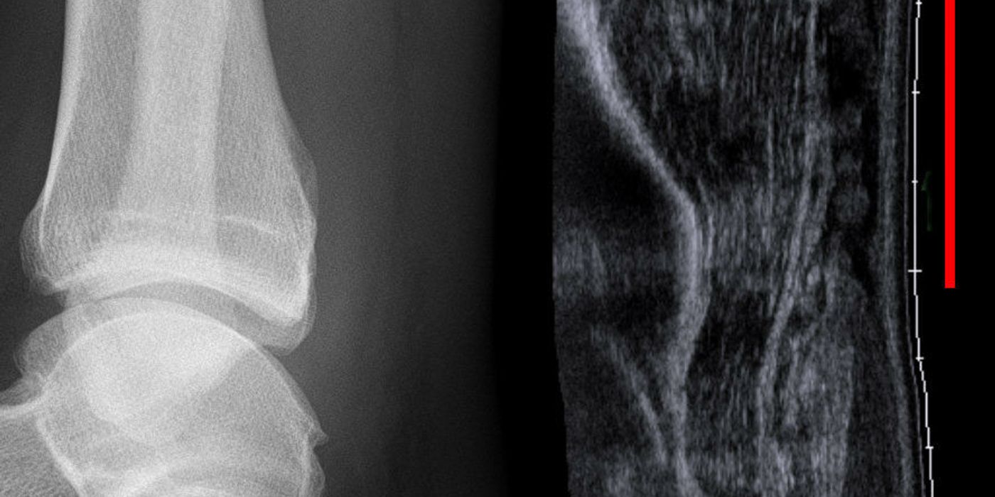
An Achilles tendon rupture is a complete tear of the calcaneal (Achilles) tendon, most often occurring 2–6 cm above its calcaneal insertion during forceful push-off, classically in adults aged 30–50 returning to intermittent sport. The diagnosis is clinical, made with the Simmonds calf-squeeze test, and in current UK practice most acute ruptures are managed non-operatively with early weight-bearing in a functional brace, which randomised trials show delivers patient-reported outcomes equivalent to surgery while avoiding wound complications.
What is an Achilles tendon rupture?
The Achilles is the conjoined tendon of gastrocnemius and soleus and the strongest tendon in the body. Rupture typically occurs in a relatively hypovascular "watershed" zone 2–6 cm proximal to the insertion, usually through a tendon already affected by degenerative tendinopathy. The classic mechanism is sudden eccentric loading — pushing off, lunging or landing — and patients frequently describe an audible snap and the sensation of being kicked or struck in the back of the leg. Recognised risk factors include pre-existing tendinopathy, fluoroquinolone antibiotics, local or systemic corticosteroids, and the deconditioned "weekend warrior" pattern of activity. Men are affected more often than women.
How is an Achilles tendon rupture diagnosed?
The diagnosis is clinical. A significant minority of ruptures are missed at first presentation, usually because the patient can still actively plantarflex using tibialis posterior, the long toe flexors and the peronei — active plantarflexion does not exclude rupture.
- Simmonds (calf-squeeze) test: with the patient prone or kneeling and the feet hanging free, squeeze the calf. Absent plantarflexion of the foot is a positive test, indicating rupture. Described by Simmonds in 1957 (and later by Thompson in 1962, hence "Simmonds–Thompson test"), it has a reported sensitivity of around 96%.
- Loss of the resting equinus: the injured foot lies in less plantarflexion than the uninjured side when both hang free (the "angle of dangle").
- Palpable gap in the tendon, usually 2–6 cm above the insertion — most reliable early, before haematoma fills the defect.
- Inability to perform a single-leg heel raise on the affected side.
Imaging is not required when the clinical diagnosis is clear. Ultrasound is useful for equivocal presentations, delayed presentations (more than about two weeks) and suspected re-rupture, where it can also assess the residual gap and apposition of tendon ends in equinus; MRI is reserved for chronic ruptures and surgical planning. This is good practice rather than the subject of a national guideline — there is currently no BOAST or NICE guideline specific to Achilles tendon rupture, so UK management is anchored to trial evidence and to NICE NG89 for thromboprophylaxis.
What is the initial management in the emergency department?
- Immobilise the ankle in full equinus — either a plaster cast in maximal comfortable plantarflexion or a walking boot with the maximum heel wedges — to appose the tendon ends. Provide crutches and analgesia.
- Refer to the next available fracture clinic or, ideally, a dedicated Achilles rupture pathway, so that definitive treatment and rehabilitation start within days.
- Perform a VTE risk assessment. As per NICE NG89 (Venous thromboembolism in over 16s), clinicians should "consider pharmacological VTE prophylaxis with low molecular weight heparin or fondaparinux sodium for people with lower limb immobilisation whose risk of VTE outweighs their risk of bleeding", and consider stopping prophylaxis if immobilisation continues beyond 42 days. Achilles rupture is a distinctly high-risk injury for VTE (see below), so most UK units offer pharmacological prophylaxis for the period of immobilisation unless contraindicated.
- Counsel the patient explicitly that both operative and non-operative treatment are evidence-based options with similar functional outcomes — this shared decision starts in the ED.
Should Achilles tendon rupture be treated operatively or non-operatively?
This is the classic interview question, and the evidence base is unusually strong.
- Willits et al., JBJS 2010: 144 patients randomised to open repair or non-operative treatment, with both arms following an accelerated functional rehabilitation protocol (early weight-bearing and early range of motion). Re-rupture occurred in 2 of 72 operative and 3 of 72 non-operative patients, with no clinically important difference in strength, range of motion or Leppilahti score, but more soft-tissue complications after surgery (13 vs 6). This trial established that when functional rehabilitation is used, the historical re-rupture advantage of surgery largely disappears.
- Ochen et al., BMJ 2019 meta-analysis: operative treatment reduced re-rupture (2.3% vs 3.9%; risk ratio 0.43, 95% CI 0.31–0.60) — an absolute risk difference of only 1.6% — at the cost of a significantly higher complication rate (4.9% vs 1.6%; risk ratio 2.76), driven mainly by infection (2.8% of operated patients).
- Myhrvold et al., NEJM 2022: 526 patients randomised three ways to non-operative treatment, open repair or minimally invasive repair. Change in Achilles tendon Total Rupture Score (ATRS) at 12 months was similar across all groups (P=0.57). Re-rupture occurred in 6.2% of non-operative patients versus 0.6% in each surgical group, and surgery carried its own risks, including nerve injury in the minimally invasive group.
Practical UK position: for an acute rupture presenting within about two weeks, non-operative management with functional rehabilitation is the default, because patient-reported outcomes are equivalent and wound complications are avoided; the trade-off is a small absolute increase in re-rupture risk (roughly 2–5 percentage points across the trials). Surgery is considered — as good practice rather than guideline mandate — for delayed presentation beyond ~2 weeks, re-rupture, a persistent tendon gap despite equinus positioning on ultrasound, and selected high-demand athletes after individualised discussion.
What does functional rehabilitation involve? The UKSTAR trial
The UK Study of Tendo Achilles Rehabilitation (UKSTAR, Costa et al., Lancet 2020) randomised 540 non-operatively managed adults across 39 UK hospitals to eight weeks in either a traditional plaster cast or a functional brace allowing immediate weight-bearing. ATRS at 9 months did not differ (adjusted mean difference −1.38, 95% CI −4.9 to 2.1); re-rupture occurred in 17 cast patients versus 13 brace patients (no significant difference); and the brace was cost-saving (−£103 per patient) with a >95% probability of cost-effectiveness at £20,000/QALY. The conclusion — early weight-bearing in a functional brace is a safe, cost-effective alternative to casting — has made bracing the standard UK pathway.
Protocols vary between units; the following is a typical example (good practice, not a national standard):
| Weeks from injury | Boot position | Weight-bearing |
|---|---|---|
| 0–2 | Full equinus (maximum wedges) | Immediate weight-bearing as tolerated in boot |
| 2–8 | Sequential wedge removal, gradually reducing plantarflexion to neutral | Full weight-bearing in boot |
| 8–10 | Wean out of boot | Physiotherapy: calf strengthening, gait re-education |
| From ~12 | — | Progressive loading; return to running/sport typically 6–9 months |
Key counselling points: the boot must be worn at all times (including in bed early on), most re-ruptures follow unprotected dorsiflexion — a slip, a stumble, or removing the boot too early — and recovery of push-off strength continues for a year or more.
What are the complications?
- Re-rupture: roughly 2–4% after surgery and 4–6% after non-operative treatment in contemporary series using functional rehabilitation (Ochen 2019; Myhrvold 2022). Manage as a new injury; many units favour operative repair for re-rupture.
- Venous thromboembolism: Achilles rupture is one of the highest-risk foot and ankle presentations. In the UK-FATE national audit (Injury, 2025), the symptomatic VTE rate after Achilles rupture was 3.69%, versus 0.57% for other foot and ankle patients. Risk-assess and treat as per NICE NG89, and maintain a low threshold for investigating calf pain or breathlessness during immobilisation — DVT symptoms are easily masked by the injury itself.
- Operative complications: wound infection and dehiscence (infection ~2.8% in the Ochen meta-analysis) in a watershed soft-tissue envelope; sural nerve injury, particularly with percutaneous and minimally invasive techniques.
- Functional deficits: tendon elongation with residual plantarflexion weakness, calf atrophy, and reduced single-leg heel-raise endurance; a minority of patients never return to their previous sporting level regardless of treatment.
Key points
- Diagnosis is clinical: positive Simmonds calf-squeeze test, palpable gap, loss of resting equinus — active plantarflexion does not exclude rupture.
- Initial management: equinus cast or fully wedged boot, VTE risk assessment (NICE NG89), and rapid referral to an Achilles pathway.
- Non-operative treatment with functional rehabilitation is the UK default: equivalent patient-reported outcomes to surgery (Willits 2010; Ochen 2019; Myhrvold 2022) with fewer complications, at the cost of a small absolute increase in re-rupture.
- UKSTAR (Lancet 2020): early weight-bearing in a functional brace is as effective as plaster casting and cost-effective — bracing is now standard.
- Consider surgery for delayed presentation (>2 weeks), re-rupture, persistent gap in equinus, and selected athletes.
- VTE risk is high (~3.7% in UK-FATE); offer pharmacological prophylaxis during immobilisation when risk outweighs bleeding risk.