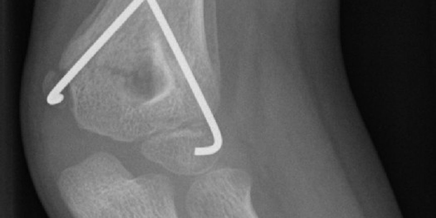
A supracondylar fracture of the humerus is a fracture of the distal humerus just above the elbow joint, through the thin metaphyseal bone between the medial and lateral columns. It is the most common elbow fracture in children, typically affecting those aged five to seven years after a fall onto an outstretched hand, and it matters because the brachial artery, and the median, radial and ulnar nerves all pass close to the fracture site. In the UK, management is defined by the British Orthopaedic Association Standard (BOAST) Supracondylar Fractures of the Humerus in Children, updated October 2020: displaced fractures are treated with closed reduction and Kirschner-wire fixation on the day of injury, with urgent surgery reserved for the ischaemic limb, open injury or threatened skin.
How are supracondylar fractures classified?
The Gartland classification (Gartland, 1959) grades extension-type fractures — the large majority, where the distal fragment displaces posteriorly — by displacement on the lateral radiograph. A multidirectionally unstable "type IV" pattern was later added (Leitch et al., 2006). Flexion-type fractures, with anterior displacement of the distal fragment, are much rarer and are classified separately.
| Gartland type | Description | Usual management |
|---|---|---|
| I | Undisplaced or minimally displaced; may only show an elbow effusion (posterior fat pad sign) | Non-operative: above-elbow cast or collar and cuff, around 3 weeks |
| II | Displaced with an intact posterior cortical hinge; anterior humeral line no longer bisects the capitellum | Closed reduction ± K-wire fixation; some minimally angulated type IIs can be managed in a cast with careful follow-up |
| III | Completely displaced, no cortical contact | Closed (or open if irreducible) reduction and K-wire fixation |
| IV | Multidirectionally unstable — the periosteal hinge is disrupted in flexion and extension (often only recognised intra-operatively) | Reduction and K-wire fixation, often with the image intensifier rotated rather than the arm |
How do children present, and what must be assessed?
The child presents with a painful, swollen elbow and reluctance to move it after a fall. S-shaped deformity, anterior bruising, skin puckering or tenting suggest significant displacement and soft-tissue interposition. The October 2020 BOAST is explicit about assessment: "a documented assessment of the limb must be performed on presentation and immediately before surgical treatment", including the radial pulse, capillary refill and the individual function of the radial, median (including anterior interosseous) and ulnar nerves. Practical points of good practice:
- Test the anterior interosseous nerve with the "OK sign" (flexion of the thumb interphalangeal and index distal interphalangeal joints) — it is the most commonly injured nerve in extension-type fractures.
- Test radial nerve motor function with wrist and finger extension, and ulnar nerve function with finger abduction; ulnar nerve palsy is more typical of flexion-type fractures.
- Document perfusion as a pulse and hand perfusion (colour, warmth, capillary refill) — the distinction drives the vascular pathway below.
Anteroposterior and lateral elbow radiographs confirm the diagnosis. On the lateral view the anterior humeral line should pass through the middle third of the capitellum; a posterior fat pad sign implies an occult fracture. Baumann's angle on the AP view helps judge coronal alignment and reduction.
When should a supracondylar fracture be operated on?
Timing is a core, examinable point of the October 2020 BOAST. The standard states: "Surgical management should be carried out on the day of injury. Night-time operating is not necessary unless there are indications for urgent surgery." Urgent surgery is defined precisely: it "should be provided urgently when there is an absent radial pulse, clinical signs of impaired perfusion of the hand and digits, open injury or evidence of threatened skin viability."
In practice this means a well-perfused Gartland III presenting in the evening is splinted in a comfortable position of around 20–30 degrees of elbow flexion (forced flexion risks vascular compromise and compartment syndrome), given analgesia, kept fasted as appropriate, and treated on a daytime trauma list the same day or the following morning — whereas the ischaemic or open injury goes to theatre immediately.
How is the pulseless limb managed — and what about the pink pulseless hand?
This is the classic viva scenario, and the BOAST resolves it. First, an absent radial pulse mandates urgent reduction in theatre — not angiography, which only delays treatment. The BOAST then states: "The majority of vascular impairments associated with supracondylar fractures resolve with fracture reduction. A limb without clinical signs of ischaemia does not require brachial artery exploration whether or not the radial pulse is present."
- Pink pulseless hand (perfused, no pulse): reduce and stabilise the fracture, then observe closely with documented serial assessment. Exploration is not required while the hand remains perfused, but any deterioration changes the pathway.
- White pulseless hand (ischaemic): a surgical emergency. Reduce the fracture urgently; per the BOAST, "if the limb remains ischaemic after fracture reduction then exploration of the brachial artery is required", with vascular or plastic surgical involvement. This is consistent with the June 2026 BOAST on arterial injuries in musculoskeletal trauma, which treats revascularisation as an NCEPOD 1 emergency and mandates consultant-led care from orthopaedics with vascular or plastic surgery.
Suspicion of compartment syndrome — pain out of proportion, pain on passive muscle stretch, escalating analgesia — "should prompt immediate vascular reassessment and intervention if required"; the July 2025 compartment syndrome BOAST requires hourly documented assessment of at-risk patients and immediate decompression once the diagnosis is made. The feared late consequence of a missed ischaemic event is Volkmann's ischaemic contracture of the forearm flexors.
What is the definitive management and which K-wire configuration should be used?
Gartland I fractures, and selected minimally displaced type IIs, are managed non-operatively in an above-elbow cast or collar and cuff for around three weeks (good practice, with early radiographic review for type II). Displaced fractures undergo closed reduction under general anaesthesia and percutaneous K-wire fixation; open reduction (usually via an anterior approach) is reserved for the irreducible fracture, open injuries and vascular exploration.
The BOAST sets the fixation standards:
- Stabilisation should use "at least two K-wires that engage in the cortex proximal to the fracture".
- "2mm diameter wires should be used, where possible, to achieve stability."
- Crossed medial–lateral wires improve stability against redisplacement, but a medial entry point risks iatrogenic ulnar nerve injury; two or three divergent lateral-entry wires avoid this. The BOAST requires that "when a medial wire is used, techniques to avoid ulnar nerve injury should be employed and recorded on the operation note" — in practice a small incision, extending the elbow while inserting the wire, and protecting the nerve.
Wires are left proud of the skin, the arm protected in a backslab, and — per the BOAST — the operating surgeon "should determine and document the need for post-operative radiographs and anticipated time of wire removal", conventionally at three to four weeks in clinic without anaesthetic (good practice).
What about nerve injuries?
Traumatic nerve injuries accompany a significant minority of displaced fractures: anterior interosseous nerve most often in extension types, radial nerve with posteromedial displacement, and ulnar nerve in flexion types. Most are neurapraxias that recover over weeks to months with observation — labelled here as accepted practice rather than a BOAST standard. The December 2021 BOAST on peripheral nerve injury requires a documented examination of all functions of the relevant nerves at first presentation and again "pre-operatively, by the operating surgeon prior to any procedure where nerve injury is a recognised risk" — precisely so that a post-operative deficit can be attributed correctly. A new deficit after surgery, particularly ulnar nerve palsy after medial wiring, warrants early senior discussion regarding wire removal or exploration, and referral pathways to a specialist peripheral nerve injury service exist for deficits that do not behave as expected.
What are the complications and long-term outcomes?
With timely reduction and stable wiring, outcomes are excellent and the BOAST notes that "routine long-term follow up is not usually required." The complications worth rehearsing:
- Malunion — cubitus varus: the classic "gunstock" deformity from coronal malreduction or loss of position. It is largely cosmetic with little functional deficit, does not remodel reliably because it is in the plane of least movement, and is corrected, when warranted, by a lateral closing-wedge (or dome) supracondylar osteotomy. Prevention — accurate reduction confirmed with Baumann's angle and stable wiring — is the real answer.
- Vascular: brachial artery injury, compartment syndrome and Volkmann's contracture, as above.
- Nerve injury: traumatic or iatrogenic, as above.
- Other: pin-site infection (usually superficial and settles with wire removal ± antibiotics), loss of reduction with inadequate wiring, stiffness (usually transient in children) and, rarely, cubitus valgus or fishtail deformity.
Key points
- Most common paediatric elbow fracture; extension-type after a fall on the outstretched hand; graded by the Gartland classification.
- Document pulse, perfusion and individual nerve function on presentation and immediately pre-operatively (Oct 2020 BOAST).
- Operate on the day of injury; night-time surgery only for the ischaemic limb, open fracture or threatened skin.
- Pink pulseless hand: reduce, stabilise and observe — no exploration while the hand is perfused; a limb still ischaemic after reduction requires brachial artery exploration with vascular/plastics.
- Fix with at least two 2mm K-wires engaging the proximal cortex; if using a medial wire, employ and document techniques to protect the ulnar nerve.
- Malunion causes cubitus varus — a cosmetic gunstock deformity treated by corrective osteotomy; accurate initial reduction prevents it.