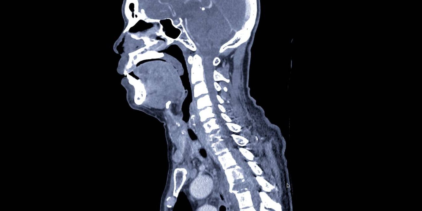
Spinal trauma assessment is the structured process of protecting, examining and imaging the spine after injury until a spinal column or spinal cord injury is either excluded ("clearing the spine") or identified and managed. In the UK it is governed by the BOAST Assessment of the Spine in the Trauma Patient (April 2025) and, where neurological deficit is present, the BOAST Management of Traumatic Spinal Cord Injury (November 2022). Traumatic spinal cord injury (SCI) is rare but devastating, and early protocol-driven care — spinal precautions, urgent CT/MRI, ISNCSCI documentation within 2 hours, and referral to a Spinal Cord Injury Centre within 24 hours — measurably reduces complications and improves outcome.
How is the spine assessed and cleared after trauma?
The April 2025 BOAST requires that "a spinal immobilisation protocol (including collars, log rolling and patient transfers) must be in place across all trauma networks", and that every trauma patient is presumed to have a spinal injury — precautions continue until clinical assessment and imaging have excluded one. Practically:
- Protect first. Apply spinal precautions (collar, in-line immobilisation, log roll for transfers) from the scene and maintain them until the spine is cleared.
- Assess clinically and document red flags: age, comorbidities, mechanism of injury, pain on movement, restricted cervical rotation and any neurological deficit. Clinical decision tools such as the Canadian C-spine rule (endorsed in NICE guideline NG41, Spinal injury: assessment and initial management) select patients who need imaging; low-risk patients who can actively rotate the neck 45° each way may be cleared clinically.
- Image with multidetector CT as the first-line modality (0.5 mm slices for the cervical spine, 0.5–1.0 mm elsewhere). Blunt cervical spinal injury should prompt CT angiography to look for vertebral artery injury.
- Use MRI when CT is contraindicated or ambiguous, in unconscious patients where ligamentous injury cannot be excluded, and whenever there are neurological symptoms or suspected cord injury.
- Report promptly: the standard specifies an initial imaging report within one hour and a definitive report within 24 hours of injury.
- Abnormal findings: if general or neurological assessment is abnormal, continue precautions and discuss with the regional spinal service.
A specific trap — clinically and at interview — is the ankylosed spine (ankylosing spondylitis, DISH). These patients fracture through fused segments after trivial trauma, the injury is unstable, and forcing the neck into a "neutral" collar position can cause or complete a cord injury. The BOAST is explicit: immobilise patients with cervical kyphosis in their own usual spinal position, and maintain a low threshold for whole-spine CT.
How are spinal injuries classified?
The AO Spine classification is the contemporary system for thoracolumbar and subaxial cervical injuries. Each injury is coded by morphology, neurological status and clinical modifiers:
- Type A — compression injuries of the vertebral body: A1 single-endplate wedge, A2 split/pincer, A3 incomplete burst (one endplate plus posterior wall), A4 complete burst (both endplates).
- Type B — tension band (distraction) injuries without translation: B1 purely osseous posterior band injury (Chance-type), B2 osteoligamentous posterior band disruption, B3 anterior tension band (hyperextension) injury — the classic ankylosing spondylitis pattern.
- Type C — translational injuries: displacement or dislocation of one vertebral level relative to another in any plane; unstable by definition.
- Modifiers: M1 indeterminate tension band integrity; M2 patient-specific comorbidity affecting treatment (e.g. ankylosing spondylitis, osteoporosis, burns over the surgical approach).
The conceptual gradient — A to C represents increasing instability, with neurological deficit escalating urgency at any morphology — is the useful message: type A injuries with an intact tension band and no deficit are usually managed non-operatively, whereas type B and C injuries generally need stabilisation. Where non-operative treatment is chosen, the April 2025 BOAST requires the presumed stability of the fracture and the management plan to be explicitly documented, with clear follow-up arrangements given to the patient.
What is the difference between spinal shock and neurogenic shock?
These are distinct entities that are frequently confused, and separating them is a reliable interview discriminator.
| Spinal shock | Neurogenic shock | |
|---|---|---|
| Nature | Transient neurological phenomenon | Haemodynamic phenomenon |
| Mechanism | Temporary loss of all cord function below the injury | Loss of sympathetic outflow (typically lesions at or above T6) with unopposed vagal tone |
| Findings | Flaccid paralysis, areflexia, absent bulbocavernosus reflex | Hypotension with bradycardia, warm well-perfused peripheries |
| Course | Resolves over roughly 24–72 hours; return of the bulbocavernosus reflex marks its end, allowing a valid assessment of injury completeness | May persist for days to weeks; managed with judicious fluids, vasopressors and atropine/pacing for bradycardia |
Two practical corollaries. First, hypotension in a trauma patient is haemorrhagic until proven otherwise — neurogenic shock is a diagnosis of exclusion. Second, the ASIA Impairment Scale grade (complete versus incomplete) cannot be reliably assigned until spinal shock has resolved. As good practice drawn from North American (AANS/CNS) guidance rather than a BOAST standard, many UK units target a mean arterial pressure of 85–90 mmHg for the first week after SCI to maintain cord perfusion.
How is traumatic spinal cord injury managed?
The November 2022 BOAST sets 12 standards for the acute phase. The ones that change what you do on the ward and in the interview answer:
- Network and escalation: every receiving hospital must have a named linked Spinal Cord Injury Centre and Specialised Spinal Surgery Centre, with 24-hour consultant access and 24-hour CT and MRI. Whole-spine MRI should follow the initial trauma CT.
- Neurological documentation: a "full detailed neurological examination should be recorded on an ISNCSCI chart, within 2 hours of admission", repeated weekly and before and after any major intervention, by clinicians trained in its use; a generalised neurological review is recorded daily.
- Systems care from day one: agreed protocols for skin (pressure area) care, gastric protection, bowel and bladder management (early catheterisation, planned bowel regimen), neuroprotection and joint care, aligned with the linked SCI Centre and national guidance.
- Referral and transfer: all patients in England are submitted to the National Spinal Cord Injuries Database within 24 hours; a joint management plan is formulated within 72 hours; and "transfer to a SCI Centre should take place within 24 hours, unless it is in the patient's best interest to remain locally".
- Surgery: anaesthetic and stabilisation protocols must follow national guidance. Regarding timing, the STASCIS study (Fehlings et al., PLoS ONE 2012) — a prospective multicentre cohort of cervical SCI — compared decompression before versus after 24 hours and underpins the "time is spine" principle of early surgical decompression in cord injury with ongoing compression, particularly incomplete injuries.
- What not to do: high-dose methylprednisolone (the NASCIS protocol) is not UK practice — NICE guideline NG41 advises against corticosteroids for acute spinal cord injury, reflecting the unfavourable balance of benefit and harm.
- Psychology: psychological support for the patient, family and carers is a named standard, not an optional extra.
What are the complications and prognosis?
Early complications include respiratory failure (diaphragmatic compromise in lesions above C5, poor cough below), neurogenic shock, pressure ulcers (preventable — hence the skin-care standard), urinary retention and paralytic ileus. Venous thromboembolism risk is very high; mechanical and, once haemorrhage risk allows, pharmacological prophylaxis should be started per local protocol (good practice). Later problems include autonomic dysreflexia in lesions at or above T6 — a hypertensive emergency triggered by noxious stimuli below the lesion, classically a blocked catheter — spasticity, syringomyelia, heterotopic ossification and chronic pain.
Prognosis is driven by injury completeness on the ASIA Impairment Scale once spinal shock has resolved: AIS A (complete) injuries rarely regain useful function below the lesion, while incomplete injuries — particularly central cord and Brown-Séquard patterns — carry meaningful potential for recovery. Sacral sparing (perianal sensation, voluntary anal contraction) is the key examination finding that reclassifies an apparently complete injury as incomplete.
Key points
- Presume spinal injury in every trauma patient; maintain precautions until the spine is cleared clinically and, where indicated, with fine-slice CT (April 2025 BOAST).
- Immobilise the ankylosed spine in the patient's own position, never forced neutral, and CT the whole spine.
- AO classification: A = compression, B = tension band failure, C = translation; instability and neurology drive surgical decision-making.
- Spinal shock is neurological and transient; neurogenic shock is haemodynamic (hypotension + bradycardia) — and hypotension is haemorrhage until proven otherwise.
- SCI care (November 2022 BOAST): ISNCSCI chart within 2 hours, database entry within 24 hours, management plan within 72 hours, transfer to an SCI Centre within 24 hours.
- Early decompression ("time is spine") for cord compression with deficit; no methylprednisolone in UK practice (NICE NG41).