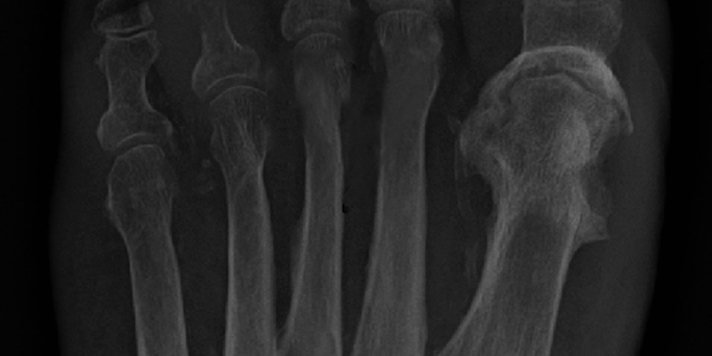
Fracture-related infection (FRI) is infection occurring at the site of a fracture, with or without metalwork in situ, and is one of the most challenging complications in orthopaedic trauma. It is diagnosed using an internationally agreed consensus definition based on confirmatory criteria (sinus, pus, matched deep cultures or positive histology), and is managed by a multidisciplinary team combining surgical debridement, fracture stabilisation, dead-space and soft-tissue management, and culture-specific antibiotics. Chronic osteomyelitis is the end-stage of untreated or failed treatment: established, often relapsing bone infection characterised by dead bone (sequestrum) that antibiotics alone cannot cure.
What is a fracture-related infection?
The term "fracture-related infection" was standardised by an international expert group (Metsemakers et al., Injury 2018, supported by the AO Foundation and EBJIS) to replace inconsistent labels such as "osteomyelitis", "infected non-union" and "deep surgical site infection" in the trauma setting. The consensus definition separates confirmatory criteria, any one of which establishes the diagnosis, from suggestive criteria, which mandate further investigation.
Confirmatory criteria:
- Fistula, sinus or wound breakdown communicating with the bone or implant
- Purulent drainage from the wound, or pus found at surgery
- Phenotypically indistinguishable pathogens cultured from at least two separate deep tissue or implant specimens
- Microorganisms in deep tissue taken at operation, confirmed on histopathological examination
Suggestive criteria include local signs (redness, swelling, warmth, pain, effusion), fever, new-onset wound drainage, raised inflammatory markers, radiological signs (loosening, sequestrum, non-union) and a single positive deep culture — the last present in the original 2018 consensus definition (Metsemakers et al., Injury 2018). FRI is conventionally divided by time from surgery — the Willenegger–Roth banding cited in the FRI consensus literature — into early (under 2 weeks), delayed (3–10 weeks) and late/chronic (beyond 10 weeks); the biologically important distinction is whether a mature biofilm has formed on implant and dead bone, which makes cure without surgery unlikely.
How is fracture-related infection diagnosed?
The Sep 2019 BOAST on Fracture Related Infections sets the UK standard. A patient with systemic sepsis and a suspected FRI needs immediate medical assessment with blood cultures, and parenteral antibiotics must not be delayed. Crucially, for the patient who is not systemically unwell, the BOAST requires consultant review within 48 hours before antibiotics are started, and states that "empiric antibiotics without a diagnostic work up should not be given" — blind antibiotics sterilise cultures, delay the diagnosis and select resistant organisms without clearing biofilm.
The diagnostic work-up per the 2019 BOAST comprises:
- Blood cultures in febrile or septic patients, plus inflammatory markers
- Plain radiographs to assess implant loosening, bone changes, sequestrum and union; cross-sectional imaging (CT for sequestrum/union, MRI for marrow involvement) as directed by the MDT
- Clinical photography of the wound with comparison against previous records
- At operation, "5 samples should be taken from around the fracture site for microbiological culture", each with clean instruments to avoid cross-contamination, plus 2 samples for histology in chronic cases
- In non-acute cases, an antibiotic-free window of at least 2 weeks before sampling
The same sampling discipline applies to any secondary fracture surgery (for example, revision of a non-union): the BOAST requires 5 microbiology and 2 histology samples unless the fracture is soundly united with no evidence of infection, because a proportion of "aseptic" non-unions prove to be low-grade FRIs.
How is early or acute FRI managed?
Management is consultant-directed with a documented plan covering sampling, debridement, fracture stability and soft-tissue cover. After deep sampling, broad-spectrum empirical antibiotics are started and reviewed at 48 hours; the BOAST states that "antimicrobial therapy should be narrowed and be culture specific as soon as possible", directed by an infection specialist with 24-hour microbiology support. All inpatients with proven or suspected FRI should have "weekly bone and joint infection MDT review".
The surgical strategy in early FRI is the fracture analogue of DAIR (debridement, antibiotics and implant retention). Retention of the implant is reasonable — this reflects the international expert-group recommendations (Metsemakers et al., Archives of Orthopaedic and Trauma Surgery 2020) rather than a numbered BOAST standard — when:
- The infection is early (typically within about 6 weeks of fixation), before a mature biofilm is established
- The implant is stable and providing satisfactory fixation of an ununited fracture
- The soft-tissue envelope is healthy or reconstructable
- A thorough excision of all infected and non-viable tissue is achievable
The key conceptual point — beloved of interviewers — is that stability suppresses infection: a stable fracture can unite in the presence of infection, whereas instability promotes it. A stable, well-fixed construct is therefore retained until union even if infection persists at a controlled level, with implant removal once the bone has healed; loose or inadequate fixation is removed and replaced (exchange fixation or external fixation), not simply retained. Suppressive antibiotics without surgery are not curative and are reserved, as a palliative or bridging strategy, for patients unfit for or declining surgery.
When is a staged strategy needed, and how are dead space and stability handled?
Late, recurrent or chronic FRI — especially with an intramedullary nail, an infected non-union or a segmental defect — usually requires implant removal or exchange with a definitive, often staged, reconstruction. The 2019 BOAST requires that these cases (late/recurrent infection, failure to respond to treatment, infected non-union and major bone defects) are discussed with an MDT focused on bone infection: orthopaedic and plastic surgeons experienced in infection management with access to a joint operating list, infection specialists and musculoskeletal radiologists — in practice a supra-regional bone infection unit, referred via the trauma network pathway.
Whatever the strategy, the same five principles apply (good practice from the international consensus literature):
- Debridement: excision of all dead bone and infected soft tissue back to bleeding, viable margins (the "paprika sign"), treating the debridement with oncological thoroughness — inadequate excision is the commonest cause of recurrence.
- Dead space management: the debridement cavity must not be left to fill with haematoma. Options include local antibiotic carriers (PMMA beads or spacers, or absorbable calcium sulphate/hydroxyapatite carriers delivering very high local antibiotic concentrations), vascularised soft tissue, and staged bone reconstruction.
- Skeletal stability: unstable fractures are stabilised, commonly with circular or monolateral external fixation in the infected field, or exchange internal fixation after adequate debridement.
- Soft-tissue cover: healthy vascularised cover, frequently a local or free flap, achieved through a combined orthoplastic approach as for open fractures (Dec 2017 Open Fractures BOAST; NICE NG37).
- Culture-specific systemic antibiotics, typically 6–12 weeks depending on strategy, rationalised by the infection team (systemic antimicrobial consensus: Depypere et al., Journal of Orthopaedic Trauma 2020).
Segmental defects after resection are reconstructed by the induced-membrane (Masquelet) technique, distraction osteogenesis/bone transport, vascularised bone grafting or, in selected cases, acute shortening; single-stage treatment is appropriate when debridement leaves a healthy, reconstructable bed, while multi-stage surgery is preferred with systemic sepsis, uncontrolled local infection or when definitive reconstruction cannot be achieved at the index procedure.
What about chronic osteomyelitis?
Chronic osteomyelitis is established bone infection with necrotic bone (sequestrum), often surrounded by reactive new bone (involucrum), draining through cloacae and sinuses, and following a relapsing–remitting course over years. It may follow FRI, open fractures, haematogenous seeding or contiguous spread (for example, diabetic foot). The Cierny–Mader staging system (Cierny, Mader and Penninck) classifies both the anatomy of the infection and the physiology of the host, and directs treatment:
| Anatomical type | Pattern | Host class | Meaning |
|---|---|---|---|
| I | Medullary (endosteal, e.g. after nailing) | A | Normal physiology |
| II | Superficial (exposed cortical surface) | B | Locally (Bl) and/or systemically (Bs) compromised |
| III | Localised full-thickness cortical sequestrum; bone stable | C | Treatment worse than the disease — suppression or palliation |
| IV | Diffuse, permeative; segmental instability |
Curative treatment follows the same principles as chronic FRI: excision of all sequestrum, dead-space management with local antimicrobials, stabilisation, soft-tissue cover and prolonged targeted antibiotics — optimising the host (glycaemic control, nutrition, smoking cessation, vascular supply) before major reconstruction. In C-hosts, long-term suppressive antibiotics or amputation may serve the patient better than limb reconstruction, and that trade-off should be an explicit, MDT-supported shared decision.
What are the complications and prognosis?
FRI risks non-union, chronic osteomyelitis, deformity, stiffness, chronic pain, amputation and, rarely, sepsis-related death; squamous cell carcinoma (Marjolin's ulcer) can arise in a long-standing sinus. Modern MDT management in specialist units achieves infection eradication in the large majority of cases, but recurrence can occur years later, so patients should be counselled that the infection is "arrested" rather than unconditionally cured. The 2019 BOAST requires every trust to run a "robust surgical site infection surveillance system" and to review re-operation rates, non-union, recurrence, amputation and death through clinical governance, and prevention remains the strongest intervention: uniform theatre and prophylaxis standards for implant surgery and management of open fractures strictly per the Open Fractures BOAST and NICE guidance.
Key points
- FRI is diagnosed by consensus confirmatory criteria: sinus/fistula, pus, ≥2 matched deep cultures, or positive histology (Metsemakers et al., 2018; diagnostic criteria updated 2020).
- Per the Sep 2019 BOAST: no empirical antibiotics without diagnostic work-up in the stable patient; consultant review within 48 hours; septic patients get cultures then immediate antibiotics.
- Deep sampling standard: 5 microbiology samples (plus 2 for histology in chronic cases), after a ≥2-week antibiotic-free window in non-acute disease — including at all secondary fracture surgery.
- Early FRI with a stable implant: debride, sample, retain fixation and treat with culture-specific antibiotics until union — stability suppresses infection.
- Late/chronic FRI and infected non-union: refer to a bone infection MDT for staged treatment — radical debridement, dead-space management with local antibiotic carriers, stabilisation, orthoplastic soft-tissue cover, then targeted systemic antibiotics.
- Chronic osteomyelitis is staged by Cierny–Mader (anatomy plus host); C-hosts may be better served by suppression or amputation than reconstruction.