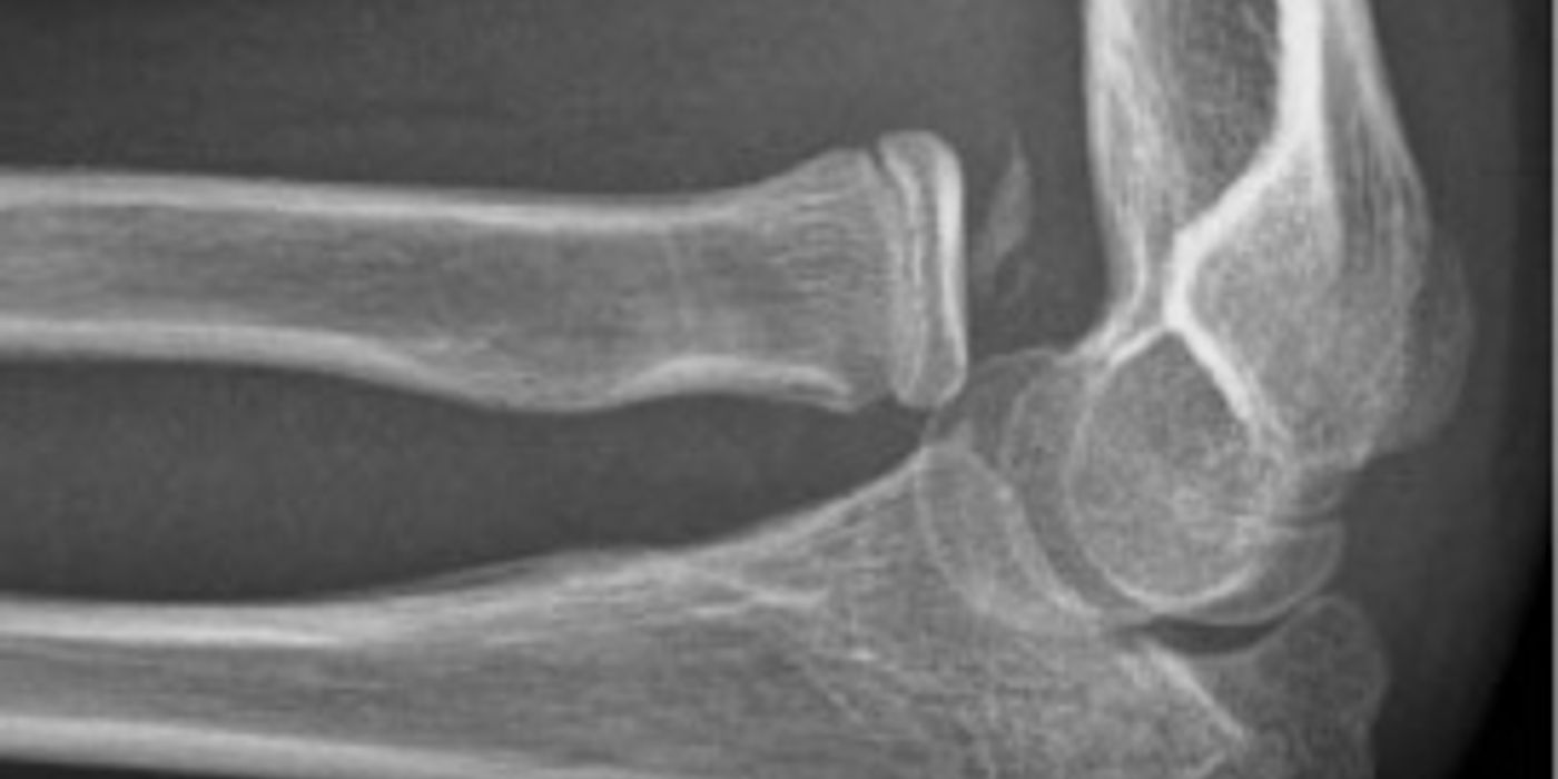
Elbow injuries in adults span a spectrum from isolated, stable radial head fractures that need almost no treatment, to fracture-dislocations — the "terrible triad" — that threaten permanent instability and stiffness. The unifying principles are: restore a concentrically reduced, stable joint; fix or replace what is needed to achieve that and no more; and mobilise early, because the elbow's chief enemy after trauma is stiffness rather than instability. This article covers the assessment and management of elbow dislocation (simple and complex), radial head fractures, olecranon fractures, the terrible triad, and the complications of stiffness and heterotopic ossification, anchored to UK standards and the trial evidence.
How should an acute elbow injury be assessed?
Assessment follows the standard trauma sequence: mechanism and energy, deformity, skin integrity, and a documented neurovascular examination of the median (including anterior interosseous), ulnar and radial (including posterior interosseous) nerves plus radial pulse and distal perfusion. As per the Dec 2021 BOAST on peripheral nerve injury, examination must be performed "at the first opportunity after injury" and repeated "after any intervention to the limb" — in practice, before and after every reduction — and documented in enough detail that sequential examinations can be compared.
- Open wounds: manage under the Dec 2017 BOA/BAPRAS Open Fracture standard — intravenous antibiotics "as soon as possible, ideally within 1 hour of injury", photograph, saline-soaked dressing, splint, and debridement within 12 hours for solitary high-energy injuries (24 hours for low-energy), planned as a combined orthoplastic case where soft-tissue cover is needed.
- Swollen, tense forearm: think compartment syndrome. The Jul 2025 BOAST highlights "pain out of proportion to the associated injury and pain on passive movement of the muscles of the involved compartments"; at-risk patients need hourly documented assessment, and diagnosis mandates immediate surgical decompression.
- Imaging: AP and lateral radiographs first; look for the posterior fat pad sign (occult fracture), congruency of the ulnohumeral joint, and the radiocapitellar line. CT is good practice for any fracture-dislocation, comminuted radial head or coronoid fracture, as it changes the operative plan.
What is the difference between a simple and a complex elbow dislocation?
A simple dislocation is a dislocation without fracture; a complex dislocation is one with an associated fracture (radial head, coronoid, olecranon or distal humerus). The distinction drives management. Most dislocations are posterolateral: the injury propagates from lateral to medial (O'Driscoll's sequence), tearing the lateral ulnar collateral ligament (LUCL) first, so residual posterolateral rotatory instability is the pattern to test for after reduction.
Simple dislocation: prompt closed reduction under sedation, neurovascular reassessment, and a post-reduction radiograph confirming a concentric joint. Examine stability through an arc of flexion-extension. The FuncSiE multicentre randomised trial (Iordens et al., British Journal of Sports Medicine, 2017; 100 patients) showed early active mobilisation was safe compared with three weeks in plaster: identical QuickDASH at one year, but less disability and a larger arc at six weeks, return to work about eight days sooner, and no recurrent dislocations. UK practice accordingly favours a sling for comfort and early active movement within the stable arc, avoiding prolonged immobilisation. Surgery is reserved for the rare elbow that will not stay reduced or re-dislocates in extension.
Complex dislocation: reduce urgently, splint, obtain CT, and plan surgery to restore bony and ligamentous stability (see terrible triad below). An irreducible dislocation, an open injury, or a new nerve deficit escalates urgency.
How are radial head fractures classified and managed?
The Mason classification (1954), with Johnston's addition of a fracture-dislocation type, remains the working language; Hotchkiss's modification ties type II to a mechanical block, which is the practical surgical trigger.
| Mason type | Definition | Management |
|---|---|---|
| I | Undisplaced or minimally displaced | Sling for comfort, early mobilisation; aspirate haemarthrosis for pain relief if needed |
| II | Displaced (>2 mm) partial articular fracture | Non-operative if no mechanical block; ORIF if a block to pronation/supination or forearm rotation |
| III | Comminuted, whole-head fracture | ORIF if reconstructible; radial head replacement if not; excision only in the low-demand, stable elbow |
| IV (Johnston) | Any radial head fracture with elbow dislocation | Treat as a complex instability pattern — fix or replace, never simply excise |
Under anaesthesia or good analgesia, test rotation: a true mechanical block, not pain, is the indication for surgery in type II injuries. In any unstable elbow the radial head is a critical secondary stabiliser against valgus and posterolateral stress, so isolated excision is contraindicated — reconstruct or replace it (good practice, consistent with the 2024 EFORT Open Reviews summary of surgical indications).
What is the terrible triad and how is it managed?
The terrible triad is an elbow dislocation with fracture of both the radial head and the coronoid process, with rupture of the LUCL (and often the MCL). It earned its name from historically poor results — recurrent instability, stiffness and arthrosis — when components were left unaddressed.
Management is operative in almost all cases, following a stepwise protocol (good practice; there is no single proven superior sequence):
- Closed reduction and splintage acutely; CT to define the coronoid and radial head fragments.
- Through a lateral (± medial or anterior) approach, working deep to superficial: fix the coronoid and anterior capsule where the fragment size warrants it.
- Radial head: ORIF if reconstructible; replacement if comminuted. Excision alone has no place here.
- LUCL repair back to the lateral epicondyle.
- Reassess stability through a full arc; if the elbow still subluxates, repair the MCL and/or apply a hinged external fixator.
- Begin protected early motion, ideally within two weeks.
A systematic review of terrible triad treatment (Chen et al., 2014) confirms that even with modern protocols complication and reoperation rates are substantial — stiffness, ulnar neuropathy, heterotopic ossification and recurrent instability — so counselling and early supervised rehabilitation matter as much as the fixation.
How are olecranon fractures managed?
The olecranon is the insertion of triceps, so displaced fractures lose active extension against gravity — the key clinical test. Management splits by displacement, comminution and patient physiology:
- Undisplaced: brief splintage then early protected motion.
- Displaced, simple transverse (younger/active): tension band wiring or plate fixation. Duckworth's randomised comparison in active adults found equivalent patient-reported outcomes but a markedly higher reoperation burden for symptomatic metalwork after tension band wiring.
- Displaced, comminuted or fracture-dislocation: plate fixation, restoring the trochlear notch.
- Elderly, low demand: non-operative management with a short period of immobilisation then mobilisation is well supported. The Edinburgh RCT (Duckworth et al., Bone & Joint Journal, 2017, patients ≥75) was stopped early because of a high complication rate in the operative arm, with no functional difference at one year; the larger SOFIE trial (Joshi et al., JBJS Am, 2025; 60 patients across 24 Australasian centres) likewise found no significant difference in DASH at 12 months and concluded non-operative treatment is "a reasonable option for displaced stable olecranon fractures in elderly patients". Patients accept an extensor lag in exchange for avoiding surgical risk.
What causes stiffness and heterotopic ossification, and what should you do about them?
Stiffness is the commonest complication of elbow trauma: the joint is highly congruent, the capsule fibroses readily, and immobilisation beyond two to three weeks predictably costs motion. A functional arc is classically 30–130° of flexion-extension and 50° each of pronation and supination. Prevention is the treatment — stable fixation that permits motion, and early supervised active mobilisation. Established stiffness that fails structured physiotherapy (± splinting) after several months may warrant open or arthroscopic capsular release, once the soft tissues are quiescent.
Heterotopic ossification (HO) — bone forming in the soft tissues — is common after fracture-dislocations, delayed surgery, repeated manipulation and head injury. Routine pharmacological prophylaxis is not supported: a Level I randomised trial of indomethacin versus placebo after surgically treated elbow trauma (164 patients, Journal of Shoulder and Elbow Surgery, 2023) found no significant reduction in HO at one year (49% vs 55%) and no difference in function. Good practice is therefore to minimise risk (single atraumatic reduction, early definitive surgery, early motion) and to excise symptomatic mature HO causing a mechanical block, typically combined with capsular release.
Key points
- Document a full neurovascular examination before and after every reduction, as per the Dec 2021 BOAST on peripheral nerve injury.
- Simple dislocations do well with reduction and early active mobilisation (FuncSiE); prolonged plaster harms function at six weeks with no benefit at one year.
- Mason II radial head fractures need surgery only for a true mechanical block; never excise the radial head in an unstable elbow.
- Terrible triad: fix the coronoid, fix or replace the radial head, repair the LUCL, then reassess — add MCL repair or a hinged fixator only if still unstable.
- Displaced olecranon fractures in low-demand patients ≥75 can be treated non-operatively (Duckworth 2017; SOFIE 2025); in the young, plates trade fewer metalwork problems against tension band wiring.
- Stiffness is the default complication — earn early motion with stable surgery; indomethacin prophylaxis for HO is not evidence-based.
- Open wounds and evolving compartment syndrome are managed to the lettered BOAST standards: antibiotics within an hour, and immediate decompression once compartment syndrome is diagnosed.