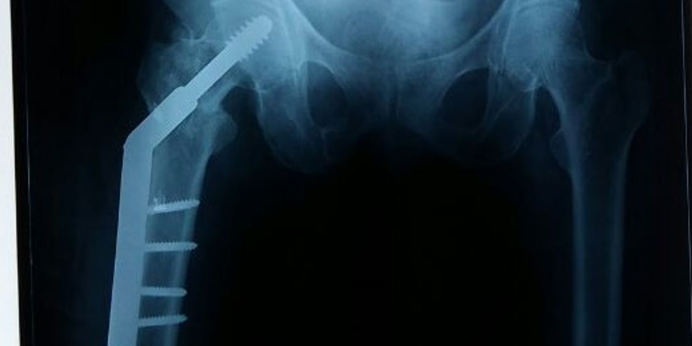
A dynamic hip screw (DHS, or sliding hip screw) is an extramedullary implant used to fix extracapsular proximal femoral fractures, most commonly stable intertrochanteric patterns. A large lag screw in the femoral head slides within the barrel of a plate fixed to the lateral femoral cortex, allowing controlled collapse and compression at the fracture site as the patient loads the limb, which promotes union while resisting varus displacement. In UK practice it is the implant recommended by NICE (CG124, Hip fracture: management) for trochanteric fractures above and including the lesser trochanter, with intramedullary nails reserved for subtrochanteric and reverse oblique patterns.
How does a dynamic hip screw work?
The DHS is a load-sharing device built around one biomechanical idea: controlled collapse. The lag screw gains purchase in the cancellous bone of the femoral head; its shaft slides freely within the barrel of a side plate that is screwed to the lateral cortex, typically at a 135° angle. When the patient weight-bears, the fracture is compressed along the axis of the barrel rather than displacing into varus or shear. The implant therefore does not need to carry the full load indefinitely — it guides the fragments into a stable, impacted position and lets the bone share load, which is why union rates in stable patterns are excellent.
Controlled collapse only works when the construct has somewhere stable to collapse to. Two structures matter:
- The medial calcar — cortical contact medially converts sliding into compression. Comminution here (AO/OTA 31A2 patterns) permits more collapse and shortening but the construct usually remains stable.
- The lateral wall — an intact lateral femoral cortex acts as the buttress against which the proximal fragment impacts. If the lateral wall is fractured or incompetent, the head–neck fragment can medialise uncontrollably past the plate and the fixation fails.
In reverse oblique and transtrochanteric fractures the fracture line parallels the direction of sliding, so "collapse" becomes medial translation of the shaft rather than compression — the mechanical reason these are contraindications.
When is a DHS the right implant — and when is it the wrong one?
NICE CG124 (Hip fracture: management) directs implant choice by fracture level:
- Trochanteric fractures above and including the lesser trochanter (AO types A1 and A2): "use extramedullary implants such as a sliding hip screw in preference to an intramedullary nail".
- Subtrochanteric fractures: "use an intramedullary nail".
Widely accepted contraindications to a DHS (good practice, consistent with the evidence below) are:
- Reverse oblique / transtrochanteric fractures (AO 31A3) — Haidukewych and colleagues (JBJS Am, 2001) reviewed 55 reverse obliquity fractures and showed sliding hip screws performed significantly worse than fixed-angle devices in this pattern.
- Subtrochanteric extension.
- A fractured or deficient lateral wall.
- Displaced intracapsular fractures in older patients, which are treated by arthroplasty rather than fixation.
A DHS with a supplementary derotation screw is an accepted fixation option for undisplaced intracapsular and basicervical fractures in patients suitable for fixation; label this as accepted practice rather than a specific NICE mandate.
| Fracture pattern (AO/OTA 31) | Preferred implant | Basis |
|---|---|---|
| A1 (simple two-part trochanteric) | DHS | NICE CG124 |
| A2 (multifragmentary, lesser trochanter involved, lateral wall intact) | DHS | NICE CG124 |
| A3 (reverse oblique / transtrochanteric) | Intramedullary nail | Haidukewych 2001; accepted practice |
| Subtrochanteric | Intramedullary nail | NICE CG124 |
What is the tip-apex distance and why does it matter?
The tip-apex distance (TAD) is the sum of the distances from the tip of the lag screw to the apex of the femoral head on the anteroposterior and lateral radiographs, corrected for magnification. It is the single best-validated predictor of cut-out — the commonest mechanical failure of the DHS, in which the screw migrates through the femoral head.
Baumgaertner and colleagues (JBJS Am, 1995) studied 198 peritrochanteric fractures: 16 fixations failed by cut-out, and the mean TAD was 24 mm in successfully treated fractures versus 38 mm in those that cut out (p = 0.0001). No screw with a TAD of 25 mm or less cut out in their series. The operative targets that follow are:
- TAD under 25 mm, checked on the intraoperative image intensifier views before reaming over the guidewire;
- centre–centre screw position in the head on both AP and lateral views — deep and central, avoiding the superoanterior quadrant.
Remember that TAD is only meaningful after an adequate closed reduction; a well-placed screw in a malreduced fracture still fails.
How is a DHS inserted, step by step?
The following sequence is standard UK technique (good practice):
- Consent, marking, WHO checklist; antibiotic prophylaxis per local policy; spinal or general anaesthesia.
- Position supine on a traction table with the injured limb in a boot and the contralateral limb positioned to allow the image intensifier to obtain true AP and lateral views.
- Closed reduction with traction and internal rotation; confirm reduction in both planes. Accept anatomical or slight valgus alignment; do not proceed on a varus reduction.
- Lateral incision from the level of the lesser trochanter distally; split fascia lata and vastus lateralis to expose the lateral femoral cortex.
- Guidewire inserted using the 135° angle guide, aiming centre–centre in the head to subchondral bone; measure length and confirm TAD <25 mm on both views.
- Triple ream over the wire (drills for the lag screw, barrel and plate seat in one pass), set to the measured length; tap in dense bone in younger patients.
- Insert the lag screw to subchondral position, ensuring the handle finishes parallel to the femoral shaft so the screw and barrel engage correctly.
- Apply the plate (commonly two or four holes) over the screw shaft, seat the barrel, and fix to the shaft with bicortical screws. Release traction to allow impaction; a compression screw may be added.
- Final image intensifier views in both planes; irrigate and close in layers.
DHS or intramedullary nail — what does the evidence say?
For stable trochanteric fractures the evidence consistently fails to show superiority of nails, while showing extra implant-related risk and cost:
- The Cochrane review by Parker and Handoll (2010; 43 trials, including 22 trials of 3,749 participants comparing the Gamma nail with the sliding hip screw) concluded that the sliding hip screw appears superior for trochanteric fractures, because cephalomedullary nails carried higher risks of intraoperative and later femoral fracture and of reoperation.
- A Swedish Hip Fracture Register cohort of 27,530 patients (Greve et al., Bone & Joint Open, 2024) found no difference between sliding hip screw and intramedullary nail in death within 120 days (hazard ratio 0.97) or in return to independent living (odds ratio 0.95) for trochanteric fractures overall.
- NICE's rationale in CG124 adds that outcomes are similar, nails have been associated with more periprosthetic fracture-related reoperation, and extramedullary implants are cheaper.
Nails retain clear roles: subtrochanteric fractures (NICE CG124), reverse oblique patterns, lateral wall incompetence, and long-segment or pathological disease. An honest interview answer is that for A1/A2 fractures the DHS is the evidence-based, cost-effective default in the UK, and that using a nail for an unstable pattern is an implant-for-fracture decision, not a preference.
What perioperative standards frame DHS surgery in the UK?
Trochanteric fracture patients are almost all older and frail, so the operation sits inside a mandated pathway. As per the May 2019 BOAST on the care of the older or frail orthopaedic trauma patient: "all surgery in the frail patient should be performed to allow full weight-bearing for activities required for daily living and within 36 hours of admission, in line with current hip fracture care", patients "should be managed in a frailty pathway which includes Comprehensive Geriatric Assessment (CGA) commencing within 72 hours of injury", and "patients should be seen by a physiotherapist on postoperative day one with early identification of functional rehabilitation goals". Practical corollaries:
- Operate on the next available trauma list; do not delay for correctable issues that can be optimised in parallel.
- Never prescribe restricted weight-bearing after a DHS — the construct is designed for immediate full weight-bearing, and frail patients cannot partially weight-bear anyway.
- Joint orthogeriatric care, delirium prevention, early mobilisation and falls/bone-health assessment complete the pathway.
What complications should you discuss?
- Cut-out — the commonest mechanical failure; risk is driven by TAD >25 mm, poor reduction, off-centre screw position and unstable patterns. Salvage in older patients is usually conversion to arthroplasty.
- Excessive collapse and medialisation — shortening, offset loss and abductor weakness with a limp; most pronounced with calcar comminution or lateral wall failure.
- Lateral wall fracture — intraoperative or postoperative; destabilises the construct and often mandates revision to a nail or fixed-angle device.
- Non-union and implant breakage — uncommon in well-vascularised trochanteric bone but seen in unstable or malreduced fractures.
- General complications of hip fracture surgery — surgical site infection, venous thromboembolism, blood loss, medical complications of frailty, and appreciable early mortality, which is why pathway metrics (time to surgery, orthogeriatric review, mobilisation) are audited nationally through the National Hip Fracture Database.
Key points
- A DHS works by controlled collapse: the lag screw slides in the plate barrel, compressing a stable trochanteric fracture as the patient weight-bears.
- NICE CG124: sliding hip screw for trochanteric fractures above and including the lesser trochanter (A1/A2); intramedullary nail for subtrochanteric fractures.
- Reverse oblique (31A3) patterns and lateral wall incompetence are contraindications — the fracture line parallels the slide, so the shaft medialises and fixation fails.
- Aim for a tip-apex distance under 25 mm with a centre–centre screw: Baumgaertner (1995) showed mean TAD 24 mm in successes versus 38 mm in cut-outs.
- Trials and registry data show no mortality or functional advantage for nails in stable patterns, with more implant-related fracture and higher cost (Cochrane 2010; Swedish register 2024).
- Operate within 36 hours and construct for immediate full weight-bearing, as per the May 2019 BOAST on the older or frail trauma patient, within an orthogeriatric pathway.