ST3 Othopaedics

ST3 Trauma and Orthopaedic Questions

ST3 ORTHOPAEDIC INTERVIEW QUESTIONS ONLINE


ABOUT

The Online ST3 Orthopaedic Interview Question Bank features over 670 ST3 trauma and orthopaedic interview questions for you to practise. The interface is fully responsive meaning you can practise questions on your phone at work or home computer.
All questions feature explanations and frameworks to help you structure your answers and understand what is expected at interview.


Sample Clinical Station Question

Key Info: The Question Bank

Our award-winning ST3 Trauma and Orthopaedic Interview preparation question bank features over 670 questions designed to help you excel at Trauma and Orthopaedic ST3 interviews. Comprehensive explanations accompany questions and videos, images and links to evidence provide easy ways for you to learn and take your existing knowledge to the next level.

More Than Questions
Also included are HD orthopaedic anatomy spot tests, CV analysis from our experienced team and regular cases to your email inbox together with our study plan to guide your preparation.

Get Your 1st Choice Job, Not Just A Job
Our materials are designed to make you stand out and we have consistently coached the highest scoring candidates at the ST3 trauma and orthopaedic interviews over the last 4 years.

Let Us Do The Hardwork For You
Accessible on any device, from anywhere everything you need to excel at the ST3 orthopaedic interviews is included with our core technology first interface helping you learn and prepare in innovative new ways.

Close

Mark and track questions that you have completed

CLINICAL SCENARIO
You are the Orthopaedic Registrar on call and are called to A&E to see a 35 year old man who has trapped his elbow and forearm in a combine harvester.

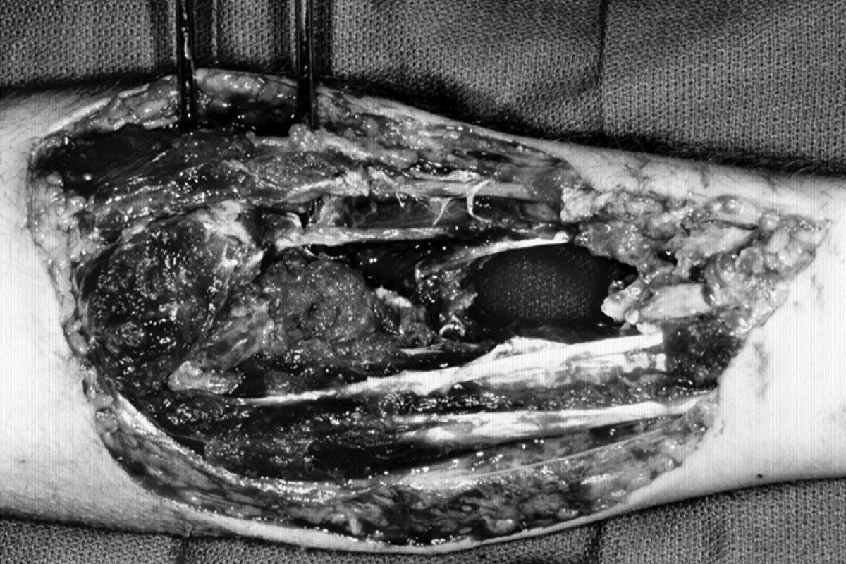
The below is an abbreviated version of a scenario from the OI ST3 Trauma and Orthopaedic Interview Questions bank.


What are the boundaries and contents of the antecubital fossa?
  • Answer

    Boundaries

    The antecubital fossa is a triangular space on the anterior aspect of the elbow bounded:
    • Superiorly: By an imaginary line between the medial and lateral condyles
    • Medially: By the lateral border of pronator teres
    • Laterally: By the medial border of brachioradialis
    • Floor: Brachialis and supinator muscles
    • Roof: Fascia and skin
    Contents
    From lateral to medial the contents can be remembered by ‘TAN’:
    • Tendon of biceps
    • Brachial artery
    • Median nerve
    • (The cephalic, basilic and median cubital veins are considered to be superficial to the fossa).
    Alternative Diagram
    Further Info
    My Image
    My Image

Describe the path of the radial nerve from the brachial plexus to the elbow
  • Answer
    The radial nerve is a branch of the posterior cord of the brachial plexus. It leaves the axilla though the triangular interval with the profunda brachii artery in the spiral groove of the humerus lying in the posterior compartment of the arm.
    It pierces the lateral intermuscular septum to leave the posterior compartment and enter the cubital fossa at the lateral epicondyle. Here it divides into the superficial radial nerve (sensory) and the posterior interosseous nerve (motor).

Describe the path of the radial nerve branches from elbow to the hand
  • Answer
    Superficial Radial Nerve
    The superficial radial nerve runs on the lateral side of the radial artery beneath brachioradialis in the forearm. Its terminal branches pass superficial to the tendons of the anatomical snuff box to supply the dorsum of the hand.

    Posterior Interosseous Nerve (PIN)
    At its origin in the cubital fossa the PIN pierces supinator 3cm distal to the head of the radius and runs in the extensor compartment of the forearm beneath the interosseous membrane to the wrist. It supplies supinator and all the forearm extensors.


How would you test the branches of the radial nerve?
  • Answer
    Sensory: skin over anatomical snuffbox (reliably supplied by superficial radial)
    Motor: wrist extension (PIN - extensors of forearm)

What does the radial nerve supply before dividing at the elbow?
  • Answer
    The radial nerve proper gives off branches to supply triceps, anconeus, brachioradialis and extensor carpi radialis longus before dividing.
    It also gives off thee sensory, cutaneous branches (posterior cutaneous nerves of arm and forearm and lateral cutaneous nerve of the arm) to supply the skin of the arm and forearm.


Describe your initial management of this gentleman
  • Answer
    This question tests your knowledge of open fracture management, BOAST guidelines and appreciation of the high energy nature of the injury.
    Key facts to mention are that this is an emergent case that is potentially limb threatening so should be treated with diligence, expedience and contacting other specialties and seniors.

    Example Answer:
    I would assess this gentleman using ATLS principles. This is a high energy injury and I would be concerned about other injuries and vascular damage leading to haemodynamic instability.

    Although it should be classified at debridement in theatre this is likely a Gustillo-Anderson III C injury and I would manage the open injury based on the BOA/BAPRAS BOAST 4 guidelines.

    After ensuring the patient is haemodynamically stable my initial management would include assessment of neurovascualr status distal to the injury. Specifically I would ensure accurate documentation of pulse, CRT and median, ulna and radial nerve examination.

    Due to the potential vascular injury I would contact the vascular surgeons, on call anaesthetist and theatres to ensure there is space to take the patient to theatre for vascular repair if required.

    I would give IV antibiotics and ensure the patient's tetanus status is up to date. I would handle the wound only to remove gross contamination, photograph the wound and then make early contact with plastic surgeons to formulate a plan for management of the soft tissues.

    I would request an X-Ray of the elbow, forearm and wrist, provide the patient with analgesia and work the patient up for theatre.”

Summary: Forearm Open Fracture

  • Clinical Summary
  • Anatomy Summary

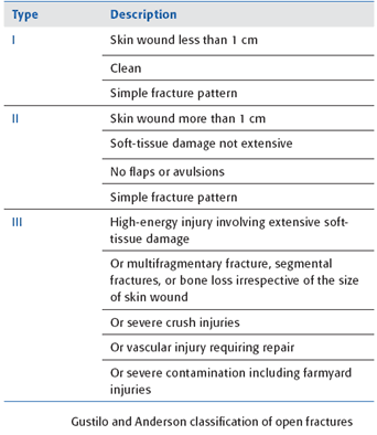
The key points from this scenario are that while the BOAST 4 guidelines are written for lower limb fractures any open fractures should be managed in a similar fashion.

Understanding that high energy injuries such as gunshots or any high energy injury automatically makes the injury a Gustillo-Anderson III.
Remember the Gustillo-Anderson classification has poor intraobserver reliability and should be used to classify at debridement.

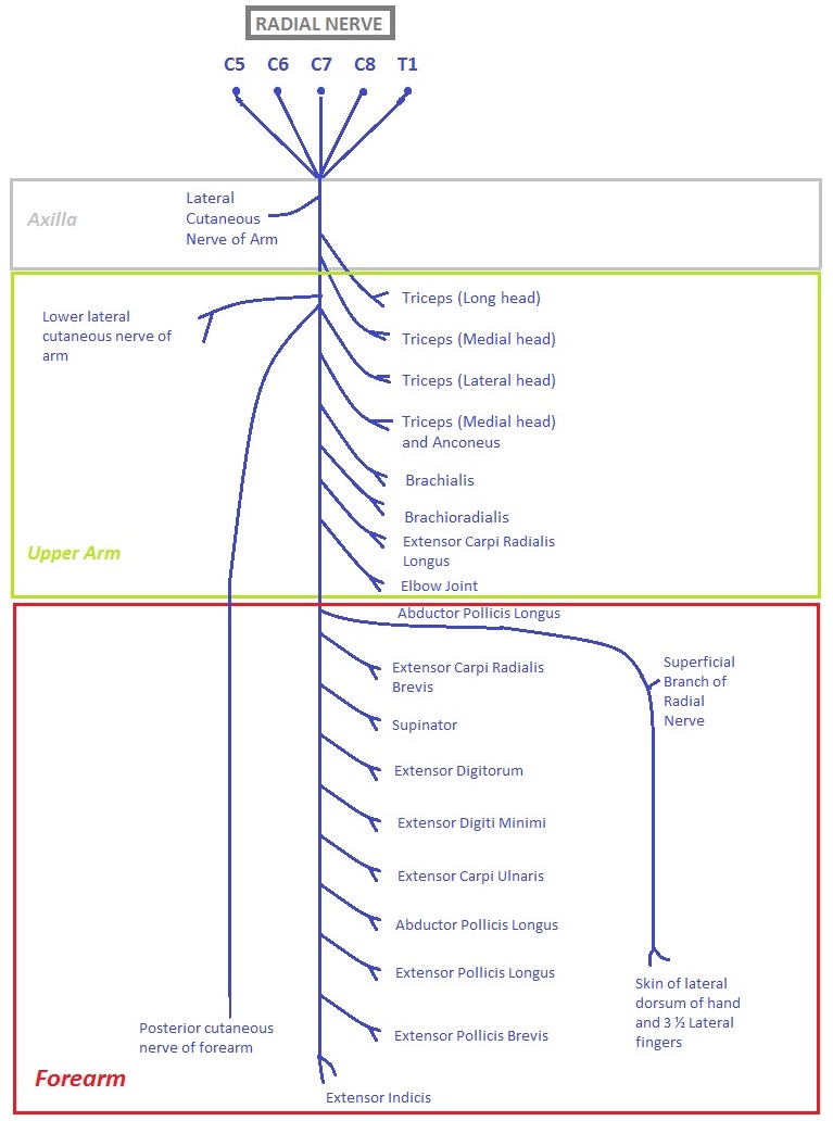
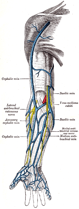
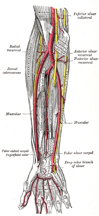
Common interview questions include the path of the peripheral nerves in the forearm, muscle attachments and what structures could be injured around the elbow and wrist.

My Image
ACF Anatomy

Boundaries

The antecubital fossa is a triangular space on the anterior aspect of the elbow bounded:
  • Superiorly: By an imaginary line between the medial and lateral condyles
  • Medially: By the lateral border of pronator teres
  • Laterally: By the medial border of brachioradialis
  • Floor: Brachialis and supinator muscles
  • Roof: Fascia and skin
Contents
From lateral to medial the contents can be remembered by ‘TAN’:
  • Tendon of biceps
  • Brachial artery
  • Median nerve
  • (The cephalic, basilic and median cubital veins are considered to be superficial to the fossa).
Alternative Diagram
Further Info
  • My Image
    ACF: Deep Structures
  • My Image
    ACF: Superficial Structures

Top Tips

  • Forearm anatomy and peripheral nerve anatomy are commonly asked topics at interview
  • Open fracture management is an easy topic to prepare for be sure you know it well
  • The OI ST3 Trauma and Orthopaedic Interview Questions Bank features more information and explanations.
  • For more questions sign up to the question bank

Further Reading
Close

CLINICAL: FEMUR ANATOMY SPOT SAMPLE

Key Info: The Question Bank

Our award-winning ST3 Trauma and Orthopaedic Interview preparation question bank features over 670 questions designed to help you excel at Trauma and Orthopaedic ST3 interviews. Comprehensive explanations accompany questions and videos, images and links to evidence provide easy ways for you to learn and take your existing knowledge to the next level.

More Than Questions
Also included are HD orthopaedic anatomy spot tests, CV analysis from our experienced team and regular cases to your email inbox together with our study plan to guide your preparation.

Get Your 1st Choice Job, Not Just A Job
Our materials are designed to make you stand out and we have consistently coached the highest scoring candidates at the ST3 trauma and orthopaedic interviews over the last 4 years.

Let Us Do The Hardwork For You
Accessible on any device, from anywhere everything you need to excel at the ST3 orthopaedic interviews is included with our core technology first interface helping you learn and prepare in innovative new ways.

Close
FEMUR ANATOMY SPOT SAMPLE
The interviewer asks you to name muscle attachments around the femur. Please complete the anatomy spot test.

Technical Skills Station: DHS Kit

TECHNICAL SKILLS SCENARIO SAMPLE: DYNAMIC HIP SCREW (DHS) KIT
The below sample questions are designed to aid you prepare for the technical skills station by reinforcing key steps and learning the kit available. This is an abbreviated version of the full question in the ST3 orthopaedic interview question bank.
Please answer the below questions regarding DHS technique

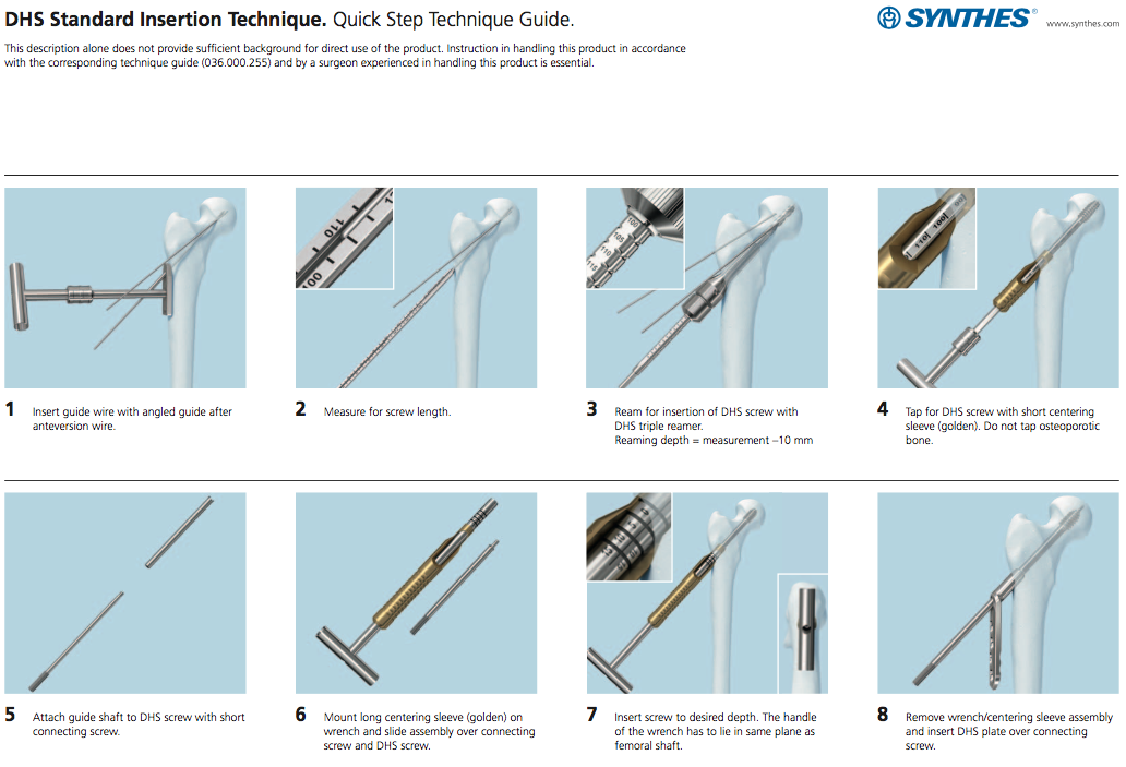
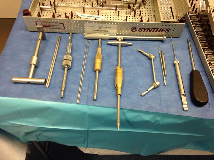
The interviewer asks you to perform a DHS on a dry bone in a clamp. You have put on gloves and an apron as suggested by the station brief. Begin by identifying the kit placed on the table before you.

Summary: Dynamic Hip Screw Kit Spot

Notes For The Interview
You will be nervous for this station and it is performed under false circumstances without image intensifier and with saw bones.

You will be asked to wear an apron and gloves.

A full explanation and summary can be found in the ST3 Trauma and Orthopaedic Question Bank.

DHS Quick Steps
Remember: Keep It Simple Stupid. The DHS really has just 5 steps:

  • Guide Wire Insertion using 135 Guide and Measure
  • Triple Reaming
  • Insert Lag Screw (+/- preceding Tap)
  • Attach Plate
  • Drill, Measure, Insert 4.5mm Plate Screws

Top Tips

  • Knowing the kit is vital for the technical skills station. In real-life a scrub nurse or senior may simply hand you the kit but being able to ask for specific kit is key for the interviews
  • The question bank features even more DHS questions together with quick look skills videos and walkthroughs to help you learn kit, steps and specifics such as size of drill bits, screws etc that might be asked at interview.

Further Reading
Close

Even More ST3 Orthopaedic Interview Questions

Mark and track questions that you have completed

EVEN MORE FREE SCENARIOS AND QUESTIONS
Follow our Facebook, Twitter and Instagram pages for even more free questions to help you prepare for the ST3 Trauma and Orthopaedic Surgery Interviews

Facebook ST3 Trauma and Orthopaedic Interview Questions


Instagram ST3 Trauma & Orthopaedic Interview Questions

{{post.caption ? post.caption.text : ''}}
View
January 1st 2017
88
Orthopaedic Interview Guides
Orthopaedic Interview Courses
Orthopaedic Interview Question Bank
Connect
NEWSLETTER

Medical imagery licensed under Creative Commons Attribution-Share Alike license; sourced from Wikipedia
All other textual content, imagery, and website design copyright © 2014-23 Ortho Interview all rights reserved.

Contact Us | Privacy Policy | Terms and Conditions